폐디스토마증(폐흡충증/폐토질/토질혈담증), Paragonimiasis
(You may visit www.drleepediatrics.com – Volume 7,
Pediatric Adolescent Infectious Diseases or제 7권, 소아 청소년 감염병 질환 웹사이트)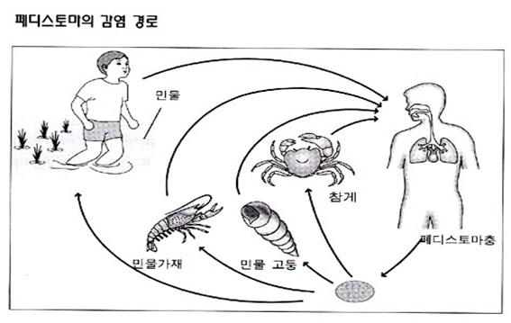
그림 5-9. 폐디스토마충에 감염되는 경로
Copyright ⓒ 2011 John Sangwon Lee, MD, FAAP
폐디스토마증(폐흡충증, 폐토질, 토질혈담증)의 원인
-
흡충류 감염을 Trematodiasis이라 한다.
-
흡충류 감염병에는 대표적으로 간디스토마증, 폐디스토마증과 장디스토마증이 있다.
-
폐디스토마증은 폐흡충(Paragonimus westermani)이나 이형폐흡충(Paragonimus heterotremus) 감염에 의해서 생긴다.
-
다시 설명하면, 폐흡충(Paragonimus westermani)이나 이형폐흡충(Paragonimus heterotremus)의 감염으로 생긴 기생충증을 폐디스토마증이라고 한다.
-
폐흡충이나 이형폐흡충을 편의상 여기서 폐디스토마충이라고 한다.
-
폐디스토마충 감염으로 생기는 기생충증을 폐디스토마증, 폐흡충증, 폐토질, 또는 토질혈담증이라고 한다.
-
폐디스토마충의 길이는 0.7∼1.2cm 정도이고, 몸통의 두께는 3∼6mm이고 수박씨 모양과 같이 납작하고 짤막하게 생겼다.
-
가래나 대변 속에 있던 폐디스토마충의 알이 민물 속에 있다가 자충으로 발육된다.
-
그 자충은 제 1 중간숙주인 다슬기에 기생하다가 제 2 중간숙주인 민물가재나 참게에 감염되어 피낭유충으로 발육된다.
-
피낭유충에 감염된 민물가재나 참게를 적절히 익혀 요리하지 않거나, 특히 날것으로 먹을 때 사람에게 감염된다.
-
경구를 통해서 인체 내로 들어온 피낭유충이 소장관 속까지 들어와서 피낭이 없어지고 새끼벌레(유충)가 된다.
-
그 유충→소장관 벽→복강→횡격막→늑막→폐 속에 도달해서 거기서 성충이 되어 기생하게 된다.
-
성충이 폐에서 기생하면서 알을 낳는다. 그 알이 기관지 속을 통과해서 가래나 침을 통해서 인체 밖으로 분비되기도 하고 그 알을 삼키기도 한다. 삼킨 알은 대변을 통해서 인체 밖으로 나올 수 있다.
-
유충에 감염된 민물가재 등을 익히지 않은 채로 먹을 때 감염될 수 있다.
-
다시 설명하면, 폐디스토마충의 새끼벌레(유충)가 오염되어 있는 민물가재나 참게 등을 생으로 먹거나 잘 익히지 않고 먹을 때, 또는 참게 젓을 담아먹을 때도 폐디스토마증에 걸릴 수 있다.
-
한국, 일본, 또는 필리핀 등 동양 여러 나라의 특수 지역에서 폐디스토마충이 발견된다.
-
과거 한 때 홍역을 생 민물가재 즙으로 치료했던 때도 있었다. 생 민물가재 즙에 있는 폐디스토마충으로 폐디스토마증에 걸려 죽기도 했다.
-
폐디스토마충의 피낭유충이 인체에 들어온 후 발병되기 전까지 잠복기는 약 8주이다.
폐디스토마증(폐흡충증/폐토질/토질혈담증)의 증상 징후
-
폐디스토마충의 유충이 감염된 민물가재나 참게를 생으로 먹은 후 피낭유충이 위장관 속으로 들어가서 탈 낭을 하고 그 때 생긴 유충이 폐 속으로 이주해서 거기서 폐디스토마증이 생긴다.
-
폐디스토마충 감염이 폐에 생기면 폐결핵을 앓을 때 생기는 증상 징후와 비슷한 증상 징후가 나타날 수 있다.
-
즉 기침·각혈·호흡곤란·숨 가쁨·가슴 아픔 등의 증상 징후가 생길 수 있고 만성기침, 늑막염(흉막염), 기흉, 기관지 확대증, 곤봉지, 폐 섬유증 등이 생길 수 있다.
-
폐디스토마충의 유충이 뇌·간·눈 또는 신체의 다른 부위로 이주해 거기서 포충 낭이 생겨 그로 인해 여러 가지의 증상 징후가 생길 수 있다.
-
폐디스토마충의 포충 낭이 안구 속에 생기면 실명할 수 있고 뇌 속에 생기면 대발작 등 전신경련을 하기도 한다.
-
폐디스토마충이 위장관 속에 기생할 때는 복통, 설사, 그 외 다른 증상들이 생길 수 있다.
-
림프절에 기생하면 림프절이 곪고 아플 수 있다.
-
폐디스토마증을 적절히 속히 치료하지 않으면 페디스토마 폐농양이 생길 수 있다.
-
폐디스토마충이 인체 내에서 약 5∼20년 간 살다가 자연히 죽는다.
폐디스토마증(폐흡충증/폐토질/토질혈담증)의 진단 치료
-
병력·증상 징후·진찰소견 등을 종합해 이 병을 진단할 수 있다.
-
민물가재나 참게를 생으로 먹은 사실과 이 병이 발병되는 지역에서 살고 있다는 정보는 이 병을 진단하는 데 큰 도움이 된다.
-
가슴 X선 사진검사로 폐디스토마충증을 진단할 수 있다.
-
안구 속에 생긴 폐디스토마 포충낭이나 피부에 생긴 폐디스토마 포충낭은 그 포충낭이 있는 조직을 통째로 떼어 조직검사로 진단할 수 있다.
-
뇌 폐디스토마 포충낭(Cysticercosis)은 뇌 CT 스캔, 뇌 MRI 검사 등으로 진단할 수 있다.
-
그러나 뇌 폐디스토마 포충낭이 있는 뇌 부위가 어디에 있느냐에 따라 진단하기가 곤란한 때도 있다.
-
가래나 대변에 있는 폐디스토마충의 알을 검사해서 진단할 수 있다.
-
결핵 피부 반응검사로 폐결핵을 진단하는 것과 거의 같은 방법으로 폐디스토마 피부 반응검사로 폐디스토마증을 진단할 수 있다.
-
폐디스토마증을 현재 앓고 있든지 과거에 이 병에 앓았다가 완치됐을 때는 폐디스토마 피부 반응검사가 양성으로 나타나는 것이 보통이다.
-
프라지퀀텔(Praziquantel)은 이 병을 치료하는 특효약이다.
-
비티오놀(Bithionol)로 치료하면 93% 정도 치료 효과가 있다.
-
그러나 이런 약으로 치료할 때 부작용이 많이 생기기 때문에 의사의 지시에 따라 치료를 받아야 한다(표 1-4 참조).
폐디스토마증(폐흡충증, 폐토질, 토질혈담증)의 예방
-
민물가재나 참게를 생으로 먹지도 말고 폐디스토마충의 새끼벌레가 오염된 민물을 끓이지 않고 마셔도 안 된다. 폐디스토마충이 유행하는 지역에서 사는 사람들은 생수를 꼭 끓여서 마셔야한다.
Paragonimiasis 폐디스토마증(폐흡충증, 폐토질, 토질혈담증)
Figure 5-9. Pathway of infection with Paragonimus heterotremus Copyright ⓒ 2011 John Sangwon Lee, MD, FAAP
Causes of Paragonimiasis
•There are 4 kinds of parasites such as Paragonimus heterotremus and others that cause Paragonimiasis.
• The length of the Paragonimus heterotremus is about 0.7~1.2cm, the thickness of the body is 3~6mm, and it is flat and short like a watermelon seed. • Paragonimus heterotremus eggs in sputum or feces are in fresh water and develop as spermatozoa.
• The larvae are parasitic to the first intermediate host, Seulgi, and then become infected with the second intermediate host, freshwater crayfish or blue crab, and develop into a cystic larva.
• Humans are infected when freshwater crayfish or sesame crab infected with metacercariae are not cooked properly, especially when eaten raw.
• The cyst larva, which has entered the human body through oral administration, enters into the small intestine, and the capsule disappears and becomes a baby worm (larva).
• The larva → the wall of the small intestine → the abdominal cavity → the diaphragm → the pleura → the lungs, where it becomes an adult and becomes a parasite.
• Adult parasites lay eggs as they parasitize in the lungs.
The egg passes through the bronchi, is secreted from the body through phlegm or saliva, and sometimes swallows the egg. Swallowed eggs can come out of the body through feces.
• Freshwater crayfish infected with larvae can be infected when eaten raw.
• In other words, when you eat freshwater crayfish or sesame crab that are contaminated with young worms (larvae) of lung Paragonimiasis, you can get Paragonimus heterotremus when eating raw, poorly cooked, or with salted sesame crab.
• Paragonimus heterotremus parasites are found in special areas in many Asian countries, such as Korea, Japan, and the Philippines.
• In the past, measles was sometimes treated with freshwater crayfish juice. He suffered from pulmonary anastomosis and died from Paragonimiasis in freshwater crayfish juice.
• The incubation period is about 8 weeks before the onset of the pulmonary cyst larvae enters the human body.
Symptoms and signs of Paragonimiasis
• After Paragonimus heterotremus larvae can be infected by eating infected freshwater crayfish or blue crab raw, the cyst larva enters the gastrointestinal tract and dislocates, and the resulting larva migrates into the lungs, causing pulmonary dystrophy there.
• Paragonimus heterotremus infections in the lungs may cause symptoms similar to those of pulmonary tuberculosis.
• In other words, symptoms such as cough, hemoptysis, shortness of breath, shortness of breath, and chest pain may occur, and chronic cough, pleurisy (pleurisy), pneumothorax, bronchiectasis, clubs, and pulmonary fibrosis may occur.
• Paragonimus heterotremus larvae migrate to the brain, liver, eyes, or other parts of the body, where they form an ensemble sac, which can lead to a number of symptomatic signs.
• If the cyst of Paragonimus heterotremus is formed in the eyeball, blindness can occur, and if it occurs in the brain, it may cause systemic convulsions such as major seizures.
• When lung Paragonimiasis parasites in the gastrointestinal tract, abdominal pain, diarrhea, and other symptoms may occur.
• Parasites in the lymph nodes can cause the lymph nodes to fester and become sore.
• If Paragonimiasis is not treated promptly and adequately, a lung abscess can occur.
• Paragonimusheterotremus live in the human body for about 5 to 20 years and then die naturally.
Diagnosis and Teatment of Paragonimiasis
• You can diagnose this disease by combining your medical history, symptoms, and examination findings.
• The fact that freshwater crayfish or crab is eaten raw and that you live in an area where the disease develops is very helpful in diagnosing the disease.
• Paragonimiasis can be diagnosed with chest X-ray examination.
• Paragonimiasis vesicles in the eyeball or Paragonimiasis vesicles in the skin can be diagnosed by biopsy by removing the entire tissues containing the parasite.
Cerebral Paragonimiasis
Cysticercosis can be diagnosed with a CT scan of the brain or an MRI of the brain.
• However, there are times when it is difficult to diagnose, depending on where the brain Cysticercosis are located in the brain.
• It can be diagnosed by examining the eggs of the Paragonimus heterotremus in sputum or feces.
• Paragonimiasis can be diagnosed with a Paragonimus heterotremus skin test in almost the same way as pulmonary tuberculosis is diagnosed with a tuberculosis skin test.
• It is common for a Paragonimus heterotremus skin test to be positive when a person has Paragonimus heterotremus or has suffered from the disease in the past and is cured.
• Praziquantel is a special drug that treats this disease.
• Treatment with Bithionol is 93% effective.
• However, many side effects occur when treated with these drugs, so you should follow your doctor’s instructions (see Table 1-4).
Prevention of Paragonimiasis
• Do not eat freshwater crayfish or blue crab raw and do not drink freshwater contaminated with Paragonimus heterotremus larvae without boiling. People living in areas where Paragonimiasis is prevalent should drink bottled water after boiling it.
출처 및 참조 문헌 Sources and references
- NelsonTextbook of Pediatrics 22ND Ed
- The Harriet Lane Handbook 22ND Ed
- Growth and development of the children
- Red Book 32nd Ed 2021-2024
- Neonatal Resuscitation, American Academy Pediatrics
- www.drleepediatrics.com제7권. 소아청소년 감염병
- Red book 29th-31st edition 2021
- Nelson Text Book of Pediatrics 19th – 21st Edition
- The Johns Hopkins Hospital, The Harriet Lane Handbook, 22nd edition
-
Childhood Emergencies in the Office, Hospital and Community, American Academy of Pediatrics
-
Emergency Medical Service for Children, By Ross Lab. May 1989. p.10
-
Emergency care, Harvey grant, and Robert Murray
-
Emergency Care Transportation of Sick and Injured American Academy of Orthopaedic Surgeons
-
Emergency Pediatrics A Guide to Ambulatory Care, Roger M. Barkin, Peter Rosen
-
Immediate care of the acutely ill and injured, Hugh E. Stephenson, Jr
-
The Critically Ill Child, Diagnosis and Management, Edited by Clement A. Smith
-
Emergency Medical Services for Children: The Role of the Primary Care Provider, America Academy of Pediatrics
-
Quick Reference To Pediatric Emergencies, Delmer J. Pascoe, M.D., Moses Grossman, M.D. with 26 contributors
-
Manual of Emergency Care
-
응급환자관리 정담미디어
-
소아가정간호백과–부모도 반의사가 되어야 한다, 이상원
-
Neonatal Resuscitation American heart Association
-
Neonatology Jeffrey J.Pomerance, C. Joan Richardson
-
Pediatric Resuscitation Pediatric Clinics of North America, Stephen M. Schexnayder, M.D.
-
Pediatric Critical Care, Pediatric Clinics of North America, James P. Orlowski, M.D.
-
Preparation for Birth. Beverly Savage and Dianna Smith
- Infectious disease of children, Saul Krugman, Samuel L Katz, Ann A. Gershon, Catherine Wilfert
-
The Harriet Lane Handbook 19th Edition
-
소아과학 대한교과서
-
제1권 소아청소년 응급의료 참조문헌과 출처
-
Other
Copyright ⓒ 2015 John Sangwon Lee, MD., FAAP
“부모도 반의사가 되어야 한다”-내용은 여러분들의 의사로부터 얻은 정보와 진료를 대신할 수 없습니다.
“The information contained in this publication should not be used as a substitute for the medical care and advice of your doctor. There may be variations in treatment that your doctor may recommend based on individual facts and circumstances. “Parental education is the best medicine.”