태변 흡인 폐렴 Meconium aspiration pneumonia
| 태변 흡인 폐렴의 개요 및 원인 |
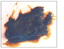
사진 163. 태변.
배안 변을 태변이라 한다.
분만이 시작 하기 전, 분만 중, 또는 분만 후 바로 태변을 볼 수 있고 그 태변이 태아의 상기도 속과 하기도 속으로 흡인될 수 있고 그렇게 해서 태어난 신생아에게 태변 흡인 폐렴이 생길 수 있다.
Copyright ⓒ 2011 John Sangwon Lee, MD., FAAP
- 태어나기 전 자궁 안에서 본 아기 변을 태변이라고 한다. 분만이 시작되기 바로 전이나 분만 중 여러 가지 원인으로 산소 공급을 충분히 받지 못할 때 태아에게 산소 결핍이 생길 수 있고 태아가 질식돼서 자궁 내에서 태변을 눌 수 있다.
- 이때 태아가 싼 태변이 양수 속에 섞이게 된다.
- 자궁 내 태아가 태변이 섞인 양수를 기도 속으로, 폐 속으로 흡인될 때 태변 흡인 폐렴에 걸리게 된다.
- 아주 묽게 섞인 양수 속 태변을 기도 속으로 흡입될 때 태아나 신생아에게 아주 경한 태변 흡인 폐렴이 생기기도 하고 전혀 생기지 않을 수 있다.
- 그렇지만 양수 속에 많이 섞인 태변이 더 많이 흡인되면 태아나 신생아에게 태변 흡인 폐렴이 더 심하게 생길 수 있다. [부모도 반의사가 되어야 한다-소아가정간호백과]-제6권 신생아 성장 발육 양호 질병-신생아의 대변, 태변 참조.
태변 흡인 폐렴의 증상 징후
- 태변 흡인성 폐렴의 증상 징후는 흡인한 태변 양과 합병증의 유무 등에 따라 다르다. 경미한 태변 흡인성 폐렴이 있을 때는 아무런 증상 징후가 생기지 않을 수 있다. 태변이 많이 섞인 짙은 양수가 흡인돼서 생긴 심한 태변 흡인성 폐렴이 생기면 다른 여러 종류의 폐렴의 증상과 거의 비슷하다.
- 즉 숨을 자주 쉬며, 신음소리를 내며, 피부가 창백하거나 파래질 수 있다. 젖이나 인공영양을 잘 빨아먹지 못할 때도 있다. 그러나 열은 나지 않는다.
태변 흡인 폐렴의 진단
- 병력, 증상 징후, 진찰소견 등을 종합해서 이 병이 의심되면 가슴 X선 사진 검사로 진단한다.
- 난산으로 태어났는지 분만 시간이 보통 이상으로 더 오래 걸렸는지 분만 전이나 분만 중 태변을 쌓는지 등에 따라 진단한다.
- 짙은 태변이 많이 섞인 양수는 멀건 푸른 콩죽과 비슷하다. 이런 태변이 기도 속으로 흡인된 후 태어난 신생아의 비강 속, 입 속, 인두 속, 기관 속에도 태변이 흡인되어 들어가 있는 것이 보통이다. 이런 이유로 다량의 태변이 흡인된 후 태어난 어떤 신생아는 숨을 제대로 쉬지 못하고 심하게 질식될 수 있다.
태변 흡인 폐렴의 치료
- 난산 분만으로 태어나거나 보통 분난 시간보다 더 오랜 분만 시간이 걸려 분만될 때는 분만 중에 태변이 양수에 섞여서 산도 속으로 나올 수 있다.
- 이런 경우 태변이 태아의 기도 속으로 흡인돼서 태변 흡인 폐렴에 걸릴 가능성이 아주 많다.
- 그래서 아기가 태어나기 전 태변이 섞인 양수가 산도를 통해 흘러나오면 아기에게 태변 흡인 폐렴이 생기지 않도록 만반의 준비를 미리 하는 것이 태변 흡인 치료의 최초 치료이다.
- 가능한 한 태변 흡인 폐렴이 생기지 않도록 치료를 효과적으로 해 주기 위해 분만 중 아기의 머리가 산도 밖으로 완전히 나오자마자 비강 속, 입 속, 인두 강 속에 있는 태변을 흡입구(사진 161)나 흡인관(사진 162)으로 흡인해내는 것이 보통이다.
- 출생 후 바로 의사가 후두경으로 검사해 인두 강 속, 후두와 기도 속, 기관 속에 태변이 있나 검진해서, 있으면 태변을 흡입구나 흡인기에 달인 흡입관으로 흡입한다.
- 이렇게 응급 치료를 해준 후 태변 흡인 폐렴이 생기는지 계속 관찰하는 것이 일반적이다.
- 숨을 자주 쉰다든지, 신음소리를 내면서 숨 쉴 때는 산소 호흡 치료를 하고,
- 포도당 전해질용액 정맥주사로 탈수를 예방치료하고,
- 몸을 보온하는 치료도 한다.
- 출생 직후 한두 시간 동안 머리와 상체를 하체보다 5도 정도 낮게 눕혀 기관 속이나 기관지 속에 있는 태변 등이 중력에 의하여 입 밖으로 흘러나오도록 치료한다.
- 몸을 이쪽저쪽으로 적절히 자주 바꿔 눕혀 기도 속에 있는 태변이 기도와 입 밖으로 흘러나오게 물리 치료한다.
- 태변 흡인 폐렴이 있을 때 찍은 가슴 X선 사진 소견은 다른 원인에 의해서 생긴 폐렴이 있을 때 찍은 가슴 X선 사진 소견과 비슷하게 나타날 수 있다.
- 따라서 신생아 박테리아 폐렴과 태변 흡인 폐렴을 서로 감별 진단하기가 곤란할 때가 많다.
- 이런 이유로 태변 흡인 폐렴이 있을 때 적절한 광범위 항생제로 우선 치료를 시작하기도 한다. 태변 흡인 폐렴이 있을 때 폐기종, 기흉, 무기폐 등의 합병증이 생길 수 있다.
- 태변 흡인성 폐렴이 심할 때는 생명이 위험할 수 있다. 폐렴 참조.
Meconium aspiration pneumonia
Overview and Causes of Meconium Aspiration Pneumonia
Photo 163. Meconium. Abdominal stool is called meconium. Meconium may be present before, during, or immediately after delivery, and the meconium may be aspirated into the upper and lower respiratory tracts of the fetus, thereby resulting in meconium aspiration pneumonia in newborns. Copyright ⓒ 2011 John Sangwon Lee, MD., FAAP
The stool seen in the womb before birth is called meconium.
Immediately before or during labor, when oxygen is not supplied enough for various reasons, the fetus may suffer from oxygen deficiency, and the fetus may suffocate and pressurize the meconium in the uterus.
At this time, the cheap meconium of the fetus is mixed in the amniotic fluid. A fetus in the womb develops meconium aspiration pneumonia when amniotic fluid mixed with meconium is aspirated into the airways and into the lungs.
When meconium in very dilute amniotic fluid is inhaled into the airways, very mild meconium aspiration pneumonia may or may not occur in the fetus or newborn. However, if more meconium mixed in amniotic fluid is aspirated, the fetus or newborn may develop more severe meconium aspiration pneumonia. [Parents should also become at least the half-doctors – Encyclopedia of Pediatric and Family Nursing] – Volume 6 Newborn Growth and Developmental Diseases – Newborn feces, refer to meconium.
Symptoms of Meconium Aspiration Pneumonia
Symptoms of meconium aspiration pneumonia vary depending on the amount of meconium aspirated and the presence or absence of complications. Mild meconium aspiration pneumonia may not cause any symptoms.
Severe meconium aspiration pneumonia caused by aspiration of thick amniotic fluid mixed with meconium is almost similar to the symptoms of other types of pneumonia. This means breathing frequently, moaning, and the skin may be pale or blue. Sometimes it is difficult to suck milk or artificial nutrition well.
But no heat.
Diagnosis of meconium aspiration pneumonia
If the disease is suspected based on the medical history, symptom signs, and examination findings, it is diagnosed by chest X-ray examination.
Diagnosis is made according to whether the child was born with difficult delivery, the delivery took longer than usual, or whether meconium was accumulated before or during delivery.
Amniotic fluid mixed with a lot of dark meconium is similar to green bean porridge. It is common for meconium to be aspirated into the nasal cavity, mouth, pharynx, and trachea of a newborn baby after this meconium is aspirated into the airway. For this reason, some newborns born after large amounts of meconium have been aspirated may not be able to breathe properly and may become severely choked.
Treatment of meconium aspiration pneumonia
If you are born with difficult delivery or deliveries that take longer than usual, meconium may mix with amniotic fluid and come out into the birth canal during delivery. In this case, the possibility of meconium aspiration pneumonia is very high because the meconium is aspirated into the fetus’ airway. Therefore, if the amniotic fluid mixed with meconium flows out through the birth canal before the baby is born, the first treatment for meconium aspiration treatment is to prepare everything in advance to prevent meconium aspiration pneumonia in the baby.
As soon as the baby’s head is completely out of the birth canal during delivery, meconium from the nasal cavity, the mouth, and the pharynx is aspirated through the suction port (Picture 161) or the suction tube (Picture 162) to provide effective treatment to avoid the occurrence of meconium aspiration pneumonia. It is normal to do Immediately after birth, a doctor examines with a laryngoscope to check if there is meconium in the pharyngeal cavity, in the larynx and airways, and in the trachea. After giving such emergency treatment, it is common to continue to observe whether meconium aspiration pneumonia develops.
When breathing frequently or breathing while moaning, administer oxygen respiration therapy, Prevention and treatment of dehydration by intravenous injection of glucose electrolyte solution, Treatment to warm the body is also performed. For an hour or two immediately after birth, the head and upper body are laid down about 5 degrees lower than the lower body, and the meconium in the trachea or bronchus flows out of the mouth by gravity.
Physiotherapy is performed so that the meconium in the airway flows out of the airway and mouth by turning the body to this side and the other side frequently.
Chest X-ray findings in the presence of meconium aspiration pneumonia may appear similar to chest X-ray findings in the presence of pneumonia caused by other causes. Therefore, it is often difficult to differentiate between neonatal bacterial pneumonia and meconium aspiration pneumonia.
For this reason, when there is meconium aspiration pneumonia, treatment with appropriate broad-spectrum antibiotics is first started.
Complications such as emphysema, pneumothorax, and atelectasis may occur when meconium aspiration pneumonia is present. In severe cases of meconium aspiration pneumonia, it can be life-threatening. See Pneumonia.
출처 및 참조 문헌 Sources and references
- NelsonTextbook of Pediatrics 22ND Ed
- The Harriet Lane Handbook 22ND Ed
- Growth and development of the children
- Red Book 32nd Ed 2021-2024
- Neonatal Resuscitation, American Academy Pediatrics
- www.drleepediatrics.com제6권 신생아 성장 발육 육아 질병
- www.drleepediatrics.com제7권 소아청소년 감염병
-
www.drleepediatrics.com제8권 소아청소년 호흡기 질환
-
Red book 29th-31st edition 2021
- Nelson Text Book of Pediatrics 19th — 21st Edition
-
The Johns Hopkins Hospital, The Harriet Lane Handbook, 22nd edition
-
Childhood Emergencies in the Office, Hospital and Community, American Academy of Pediatrics
-
Emergency Medical Service for Children, By Ross Lab. May 1989. p.10
-
Emergency care, Harvey grant, and Robert Murray
-
Emergency Care Transportation of Sick and Injured American Academy of Orthopaedic Surgeons
-
Emergency Pediatrics A Guide to Ambulatory Care, Roger M. Barkin, Peter Rosen
-
Immediate care of the acutely ill and injured, Hugh E. Stephenson, Jr
-
The Critically Ill Child, Diagnosis and Management, Edited by Clement A. Smith
-
Emergency Medical Services for Children: The Role of the Primary Care Provider, America Academy of Pediatrics
-
Quick Reference To Pediatric Emergencies, Delmer J. Pascoe, M.D., Moses Grossman, M.D. with 26 contributors
-
Manual of Emergency Care
-
응급환자관리 정담미디어
-
소아가정간호백과–부모도 반의사가 되어야 한다, 이상원
-
Neonatal Resuscitation American heart Association
-
Neonatology Jeffrey J.Pomerance, C. Joan Richardson
-
Pediatric Resuscitation Pediatric Clinics of North America, Stephen M. Schexnayder, M.D.
-
Pediatric Critical Care, Pediatric Clinics of North America, James P. Orlowski, M.D.
-
Preparation for Birth. Beverly Savage and Dianna Smith
- Infectious disease of children, Saul Krugman, Samuel L Katz, Ann A. Gershon, Catherine Wilfert
-
The Harriet Lane Handbook 19th Edition
-
소아과학 대한교과서
-
제1권 소아청소년 응급의료 참조문헌과 출처
-
Other
Copyright ⓒ 2015 John Sangwon Lee, MD., FAAP
“부모도 반의사가 되어야 한다”-내용은 여러분들의 의사로부터 얻은 정보와 진료를 대신할 수 없습니다.
“The information contained in this publication should not be used as a substitute for the medical care and advice of your doctor. There may be variations in treatment that your doctor may recommend based on individual facts and circumstances. “Parental education is the best medicine.”