외출혈과 내출혈의 응급처치 Emergency treatment for external bleeding (external hemorrhage) and internal bleeding (Internal hemorrhage)
- 몸속 한 부위에서 바로 그 주위에 있는 다른 부위 속으로 출혈하는 것을 내출혈 또는 내부 출혈이라고 하고 몸속의 피가 몸 밖으로 출혈하는 것을 외출혈 또는 외부출혈이라고 한다.
- 출혈할 때 내출혈만 할 때도 있고 외출혈만 할 때도 있고 내출혈과 외출혈을 동시 할 때도 있다.
-
내출혈이나 외출혈의 원인은 여러 가지이다.
-
혈소판 감소증, 패혈증, 혈우병, 또는 외상 등으로 내출혈만 할 수도 있고, 또 외출혈만 할 수도 있고 외출혈 및 내출혈을 동시 할 수 있다.
-
찰과상, 열상, 자상, 골절, 또는 총상 등으로 외출혈 및, 또는 내출혈을 동시에 할 수 있다.
-
여기서는 외출혈에 관해 주로 설명한다
-
급성 출혈에 의한 빈혈 참조).
외(부)출혈과 내(부)출혈의 증상 징후
-
외출혈과 내출혈의 원인과 정도에 따라 증상 징후가 다르다.
-
출혈로 전체 혈량의 15% 정도 갑자기 잃으면 쇼크에 빠질 수 있고,
-
전체 혈량의 30% 정도 삽시간에 잃으면 심한 쇼크에 빠질 수 있다.
-
출혈로 삽시간에 많은 피를 잃으면 신체 모든 조직이 필요로 하는 혈 량을 충분히 공급받을 수 없다. 따라서 신체의 모든 세포에게 산소 결핍증이 생길 수 있다.
-
혈 량이 급격히 감소되면 혈압이 정상 이하로 떨어지고 심장박동이 정상 이상 빨라질 수 있고 약해진다.
-
심장이 비정상적으로 오랫동안 빠르게 박동하면 심장이 쇠약해지고 심폐 부전증이 생길 수 있다.
-
출혈로 상당히 많은 피를 삽시간에 잃으면 불과 몇 분 내 쇼크에 빠질 수 있고 심지어는 사망하게 된다.
-
교통사고나 총상 등으로 생긴 심한 외출혈은 대개 내출혈을 동반ㅅ하는 것이 보통이다.
-
외출혈이 심할 때는 피가 나는 상처를 손으로 눌러 지혈하거나(사진204~210 참조) 다른 지혈 방법으로 응급 지혈을 한다.
-
외출혈이나 내출혈로 피를 많이 잃어 쇼크에 빠질 때는 지체하지 말고 현장에서 응급처치를 즉시하고 구급대원이나 의사 또는 병원 응급실의 도움을 청하고 받아야 한다.
-
상황에 따라 뇌 심장 폐 간 신장 등 인간 생명유지 기관들에 피가 더 많이 흘러가게 하체를 상체보다 15∼30도 정도 더 높게 눕힌다.
-
가능한 한 구급차를 이용해서 수혈치료를 받을 수 있고 그 외 다른 추적 응급치료를 적절히 받을 수 있는 큰 병원 응급실로 급히 이송 한다.
-
외출혈 세분 응급처치법을 다음 설명한다.
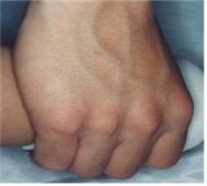
사진 194. 손등의 정맥
Copyright ⓒ 2012 John Sangwon Lee, MD., FAAP
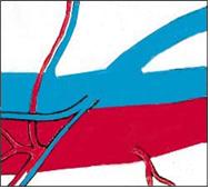
그림 195. 동맥(적색)과 정맥(청색)
혈관의 크기에 따라 다르지만 일반적으로 동맥에서 나는 피는 새빨갛고 확 솟아나는 것이 보통이고 정맥에서 나는 피는 검붉고 조금씩 솟아나거나 스며나는 것이 보통이고 모세혈관에서 나는 피는 조금씩 스며나는 것이 보통이다.
Copyright ⓒ 2012 John Sangwon Lee, MD., FAAP
출혈의 종류
1. 동맥에서 나는 외출혈과 내출혈
-
동맥에서 나는 피는 선명한 적색이고 심장의 수축과 이완 주기에 따라 피가 더 분출하거나 덜 분출하는 식의 출혈이 생기는 것이 보통이다.
-
절상이나 자상 등으로 절단된 큰 동맥에서 나는 외출혈이나 내출혈은 자연적으로 멎지 않기 때문에 그 외출혈이나 내출혈을 즉시 지혈시키지 않으면 짧은 시간에 다량 출혈해 생명이 위험할 수 있다.
-
큰 정맥이 절단될 때도 심하게 출혈될 수 있고 자연적으로 출혈이 멈추지 않지만 작은 정맥이 절단될 때는 절단된 정맥의 양 끝 부분이 자연적으로 수축되어 절단된 정맥관 끝 부분이 꼭 막힐 수 있다. 절단된 정맥 끝 부분에 혈액 응고가 생겨 자연히 지혈될 수 있다.
-
외출혈이나 내출혈로 많은 피가 짧은 시간 내 소실될 때는 삽시간에 쇼크에 빠져 사망할 수 있다.
2. 정맥에서 나는 외출혈과 내출혈
-
작은 정맥에서 나는 외출혈과 내출혈의 색은 검푸르고 심장이 수축될 때마다 피가 분출되지 않고 조금씩 계속 흘러나오는 것이 보통이다.
-
그렇지만 큰 정맥이 절단되었을 때는 동맥에서 나는 외출혈이나 내출혈과 거의 비슷하게 피가 다량으로 출혈할 수 있다.
-
일반적으로 정맥에서 나는 외출혈이나 내출혈은 동맥에서 나는 외출혈이나 내출혈보다 지혈시키기가 훨씬 더 쉽다.
-
심장 가까이 있는 큰 정맥이 절단되거나 멀리 있는 큰 정맥이 절단되면 공기나 피 덩어리가 절단된 정맥관 속으로 들어갈 수 있다. 정맥혈과 같이 심장 속으로 들어온 공기나 핏 덩어리가 폐동맥 혈관 속으로 들어갈 수 있고 공기나 핏덩어리로 폐동맥 혈관 속이 막혀서 심장에서 폐 속으로 피가 정상적으로 흐를 수 없는 때도 있다.그에 따른 여러 가지의 증상 징후가 생길 수 있다.
-
이렇게 공기로 생긴 공기 색전을 공기색전증(空氣塞栓症) 핏 덩어리로 생긴 색전을 혈색전증이라고 한다.
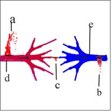
그림 196. 외출혈
a-동맥에서 나는 외출혈, b-정맥에서 나는 외출혈
c-모세혈관에서 나는 외출혈, d-동맥, e-정맥
Copyright ⓒ 2012 John Sangwon Lee, MD., FAAP
3. 모세혈관에서 나는 외출혈과 내출혈
-
피부가 얇게 벗겨지거나 손상되거나 또는 신체 내 어떤 손상이 생길 때 모세혈관이나 림프관 등에서 피와 조직액과 림프액 등이 체외 또는 그 주의 체내로 조금씩 스며 나올 수 있다.
-
선행 건강문제가 있지 않는 한 대개의 경우 모세혈관에서 나는 피는 자연적으로 지혈되는 것이 보통이다.
출혈의 진단
-
외출혈이 있을 때 증상 징후, 병력을 참작하여 육안으로 보고, 검진해서 외출혈이 있다고 바로 진단할 수 있다.
-
외출혈과 내출혈이 동시 있을 때도 병력, 검진 등으로 진단할 수 있으나 내출혈을 진단하는 데는 출혈의 정도와 출혈하는 신체의 부위에 따라 초음파 검사, X-선 검사, CT 사진 검사 등 여러 가지 검사로 진단 할 때도 있다.
-
원인을 확실히 알 수 없는 내 출혈이나 외 출혈이 생기면 출혈의 원인을 확실히 알면 치료를 효과적으로 한다.
-
또 그 출혈의 원인에 따라 치료를 달리한다.
-
출혈로 생긴 증상 징후에 따라 치료한다.
-
비 정상적으로 피가 나면 CBC 피 검사, 프로트롬빈 시간(Prothrombin time/PT), 부분적 트롬보플라스틴 시간(Partial thromboplastin time/PTT), 출혈 시간(Bleeding time) 검사 등 혈액검사로 출혈의 원인을 더 확실히 알아보고 원인에 따라 치료 한다.
-
출혈이 있을 때 의사들은 다음 표 23에 있는 여러 가지 임상검사를 분별 있게 한다.
표 23. 출혈하는 환아의 출혈 스크린 검사
| 출혈 스크린 검사 출혈이나 이상 | 유전 또는 후천적 | 혈소판 수 | 출혈시간 BT | 부분적 트롬보플라스틴 시간 Partial thromboplastin time/PTT | 프로트롬빈 시간 prothrombin time/PT | TT/Thrombin time | 참조 |
| 정상 출혈 스크린 검사치 | – | 150,000-400,000/ ml | 4~9분 | 25~35초 | 12~13초 | 8~10초 | 섬유소원 레벨 190-400 mg/dl |
| 혈우병 A-혈액응고 인자 VIII | 유전병 | 정상 | 정상 | 증가 | 정상 | 정상 | 인자분석 |
| 혈우병 B- 혈액응고 인자 IX (크리스마스B) | 유전병 | 정상 | 정상 | 증가 | 정상 | 정상 | 인자분석 |
| 혈액응고 인자 XI | 유전병 | 정상 | 정상 | 증가 | 정상 | 정상 | 인자분석 |
| 혈액 응고 인자 XII | 유전병 | 정상 | 정상 | 증가 | 정상 | 정상 | 인자분석 |
| 혈액응고 인자 II, V, X | 유전병 | 정상 | 정상 | 증가 | 증가 | 정상 | 인자분석 |
| 혈액응고 인자 VII | 유전병 | 정상 | 정상 | 정상 | 증가 | 정상 | 인자분석 |
| 폰 빌리부란트( 여러 형이 있음) | 유전병 | 정상 | 증가 | 증가 | 정상 | 정상 | 폰 블리부란트 항원과 활성등 |
| 혈소판 기능 부전 | 유전병 | 정상 또는 감소 | 증가 | 정상 | 정상 | 정상 | 혈소판 응집검사 |
| 파종성 혈관내 응고 | 후천적 질병 | 감소 | 증가 | 증가 | 증가 | 증가 | 섬유소원레벨이 감소되고, 섬유 소원 분리물이 증가 |
| 특발성 혈소판 감소성 자반 | 후천적 질병 | 감소 | 증가 | 정상 | 정상 | 정상 | 혈소판 파괴가 증가 |
| 헤느흐 쉰라인 자반(색)증 | 후천적 질병 | 감소 | 정상 | 정상 | 정상 | 정상 | – |
| 간장 부전증 (심할 때) | 후천적 질병 | 정상 또는 감소 | 정상 또는 증가 | 증가 | 증가 | 정상 또는 증가 | 섬유소원 레벨이 감소, 섬유 분리 물이 증가– |
| 요독증 | 후천적 질병 | 정상 또는 감소 | 증가 | 정상 | 정상 | 정상 또는 증가 | 이 차성 간부전증 또는 단백질 소실 |
| 헤파린 치료 | 후천적 질병 | 정상 | 정상 | 증가 | 정상 또는 증가 | 많이 증가 | – |
| 쿠마딘(Coumadin)제 치료 | 후천적 질병 | 정상 | 정상 | 정상 또는 증가 | 증가 | 정상 | – |
| 용혈성 요독 증후군, 혈전성 혈소판 감소성 자반 | 후천적 질병 | 감소 | 변화 | 정상 | 정상 | 정상 | – |
출처:Emergency Pediatrics, 5th Ed. Roger m. Barkin. Peter Rosen p.222
출혈의 응급치료
-
얼굴, 팔, 또는 다리 등에 생긴 경미한 찰과상, 자상, 또는 열상(절상)에서 나는 경미한 외출혈은 특별히 치료하지 않아도 자연히 지혈된다.
-
그러나 심한 자상이나 절상 등으로 절단된 동맥에서 나는 외출혈의 거의가 자연적으로 지혈되지 않는 것이 보통이다.
-
삽시간에 피를 다량으로 흘리면 쇼크에 빠질 수 있기 때문에 쇼크에 대한 처치를 동시 한다.
-
피가 조금 날 때도 우선 아이를 안정시키는 동시 지혈시켜야 한다.
-
신체 어느 부위가 찔리거나 찢어져 거기서 외출혈이 조금 날 때는 손가락이나 맨손으로 피가 나는 상처를 꼭 눌러 우선 지혈한다.
-
이렇게 지혈하는 방법이 가장 빠르고 가장 쉬운 응급 지혈 처지법이다.
-
가능한 한 멸균 거즈, 수건, 또는 헝겊 조각 등을 피가 나는 상처 위에 얹어놓고 그 위에 손가락 또는 손바닥을 올려놓고 출혈 상처를 꼭 눌러 지혈 처치한다.
-
자상이나 열상 등으로 큰 동맥이나 큰 정맥이 잘려서 외출혈이 심할 때는 찢어진 상처나 찔린 상처를 손으로 직접 꼭 눌러 지혈시킨다.
-
그와 동시 필요에 따라 피가 나는 상처에서부터 심장이 있는 쪽 가까운 신체 한 부위를 붕대, 허리띠, 넥타이, 또는 지혈대 등으로 꼭 매어 지혈시킬 수 있다.
-
넥타이나 지혈대 등으로 신체 일부를 일시적으로 꼭 매어 지혈시킬 때는 적어도 몇 초 마다 맨 지혈대를 잠시 동안 풀어놓았다가 다시 매는 식으로 지혈한다.
-
팔이나 다리 등이 절단되어 출혈이 심할 때에도 같은 처치방법으로 지혈할 수 있다.
-
붕대나 허리띠, 또는 지혈대 등으로 출혈되는 혈관을 압박하기 위해 신체 일부를 매서 지혈하는 방법은 다른 방법으로 지혈이 안 될 때 하는 최후 지혈 처치 방법이다.
-
이 방법으로 지혈을 해 줄 때는 출혈하는 혈관을 압박해서 맨 부위 이하 부분에 혈액 공급이 일시적으로 차단될 수 있기 때문에 그와 동시에 매어준 신체 말초부위가 손상될 수 있기 때문이다.
-
심하게 출혈될 때 쇼크를 예방하기 위하여 머리와 상체를 하체보다 5~10도 정도 낮게 눕힌다.
-
의료구급대원, 병원 응급실, 또는 의사에게 응급으로 전화상담 후 그들의 지시에 따라 응급실로 급히 데리고 가서 치료 받을 수 있다.
-
신체의 어느 부위가 절단되었을 때는 절단 신체 부위를 가능한 한 생리식염수 속에 담아가지고 환아와 같이 병원 응급실로 간다.
-
피가 심하게 날 때 피가 나는 상처를 손으로 직접 압박하고, 동시에 상처 난 부위에 피를 공급해 주는 동맥이 있는 신체 부분을 손으로 직접 압박하거나 지혈대로 매어 지혈시킬 수 있다.
-
입안 점막층, 혀, 또는 잇몸 등에서 경미하게 출혈될 때는 손가락 끝으로 출혈 상처를 가만히 눌러 지혈시킬 수 있다.
-
그 다음 거즈로 피를 닦고 출혈 상처를 살펴본다.
-
입안에서 피가 날 때도 손가락 끝으로 출혈하는 국소를 1~2분 동안 꼭 누른다. 가능하면 얼음 덩어리를 입안에 잠시 물고 있으면 경미한 출혈은 쉽게 지혈된다.
-
상처와 출혈의 정도에 따라 병원으로 아이를 데리고 가서 치료 받는다.
Emergency treatment for external bleeding (external hemorrhage) and internal bleeding (Internal hemorrhage)
외출혈과 내출혈의 응급처치
• Bleeding from one area of the body into another area around it is called internal bleeding or internal bleeding, and bleeding out of the body is called external or external bleeding.
• When bleeding, sometimes only internal bleeding occurs, sometimes only external bleeding, and sometimes both internal and external bleeding.
• There are many causes of internal or external bleeding. • You may have only internal bleeding due to thrombocytopenia, sepsis, hemophilia, or trauma, and you may only have external bleeding, or you may have both external and internal bleeding.
• External and/or internal bleeding can occur simultaneously due to abrasion, laceration, cuts, fractures, or gunshot wounds.
• This section mainly describes external bleeding.
• Anemia due to acute bleeding).
Symptoms of external bleeding and internal bleeding
• Symptoms and signs differ depending on the cause and severity of external and internal bleeding.
• Sudden loss of about 15% of total blood volume due to bleeding can lead to shock,
• If you lose about 30% of your total blood volume quickly, you can suffer from severe shock.
• If you lose a lot of blood quickly due to bleeding, you cannot get enough blood volume that all the tissues of your body need. Thus, oxygen starvation can occur in every cell of the body. • When blood volume decreases rapidly, blood pressure drops below normal and the heart rate may become faster and weaker than normal.
• If the heart beats fast for an abnormally long period of time, it may cause weakness and cardiopulmonary failure.
• If you lose a lot of blood quickly from bleeding, you can shock and even die within minutes.
• Severe external bleeding caused by traffic accidents or gunshot wounds is usually accompanied by internal bleeding.
• When external bleeding is severe, stop the bleeding by pressing the bleeding wound with your hand (see pictures 204~210), or perform emergency hemostasis using other hemostasis methods.
• If you lose a lot of blood due to external or internal bleeding and you are in shock, do not delay, take first aid at the site immediately, and seek help from an paramedic, doctor, or hospital emergency room.
• Depending on the situation, lay the lower body 15 to 30 degrees higher than the upper body so that more blood flows to the human life-supporting organs such as the brain, heart, lungs, liver, kidneys.
• As soon as possible, transfer to a large hospital emergency room where you can receive blood transfusions by ambulance and receive other follow-up emergency care as appropriate.
• The following is a detailed first aid method for external bleeding.
Picture 194. Veins on the back of the hand Copyright ⓒ 2012 John Sangwon Lee, MD., FAAP Figure
195. Arteries (red) and veins (blue) It depends on the size of the blood vessels, but in general, the blood from the arteries is usually red and bulging out, the blood from the veins is dark red, and the blood from the capillaries is usually oozing or oozing out little by little. Copyright ⓒ 2012 John Sangwon Lee, MD., FAAP
Type of bleeding
1. External and internal bleeding from arteries
• Blood from the arteries is bright red, and bleeding occurs in the form of more or less bleeding depending on the contraction and relaxation cycle of the heart.
• Because external or internal bleeding from a large artery that has been cut due to a cut or cut does not stop naturally, if the external or internal bleeding is not stopped immediately, a large amount of bleeding in a short time can be dangerous.
• When a large vein is cut, the bleeding can be severe and the bleeding does not stop naturally, but when a small vein is cut, the ends of the cut vein are naturally constricted and the ends of the cut vein can be blocked. Blood clots can form at the ends of the severed veins, which can cause hemostasis to stop naturally.
• When a lot of blood is lost within a short period of time due to external or internal bleeding, shock can occur quickly and die.
2. External and internal bleeding from veins
• The color of external bleeding and internal bleeding from small veins is dark blue. Whenever the heart contracts, the blood does not squirt, but it is common to keep bleeding out little by little.
• However, when a large vein is cut, large amounts of blood can bleed, much like external or internal bleeding from an artery. • In general, external or internal bleeding from a vein is much easier to stop bleeding than from an external or internal bleeding from an artery.
• If a large vein near the heart is cut or a large vein in the distance is cut, air or blood clots can enter the cut vein. There are times when air or blood clots that have entered the heart, such as venous blood, can enter the pulmonary artery vessels, and the blood flow from the heart to the lungs is not normal due to the blockage of the blood vessels in the pulmonary arteries with air or blood. I can.
• Air embolism caused by air in this way is called thromboembolism.
Figure 196. External bleeding a-external bleeding from arteries, b-external bleeding from veins c-capillary external bleeding, d-artery, e-vein Copyright ⓒ 2012 John Sangwon Lee, MD., FAAP
3. External and internal bleeding from capillaries
• When the skin is thinly peeled or damaged, or any damage occurs in the body, blood, tissue fluid, and lymph fluid from capillaries or lymphatic vessels may seep out little by little from the body or into the body around the body.
• Unless there is a prior health problem, blood from the capillaries usually stops naturally.
Diagnosis of bleeding
• When there is external bleeding, you can immediately diagnose that there is external bleeding by visually seeing and examining the symptoms, symptoms and medical history.
• When external bleeding and internal bleeding are at the same time, it can be diagnosed by medical history, examination, etc. However, to diagnose internal bleeding, it is diagnosed by various tests such as ultrasound, X-ray, and CT photographs, depending on the degree of bleeding and the part of the bleeding body. Sometimes I do.
• If internal or external bleeding occurs for which the cause is unknown, treatment is effective if you know the cause of the bleeding.
• Also, treatment is different depending on the cause of the bleeding.
• Treat according to the symptoms of bleeding.
• If you bleed abnormally, use blood tests such as CBC blood test, Prothrombin time/PT, Partial thromboplastin time/PTT, and bleeding time to determine the cause of bleeding more clearly. Find out and treat according to the cause.
• When bleeding occurs, doctors sensitize the different clinical tests listed in Table 23 below.
Table 23. Bleeding screen examination of children with bleeding
표 23. 출혈하는 환아의 출혈 스크린 검사
| Bleeding screen inspection Bleeding or abnormality | Hereditary or acquired | Platelet count | Bleeding time: BT | Partial thromboplastin time/PTT | prothrombin time/PT | TT/Thrombin time | Reference |
| Normal bleeding screen test value | – | 150,000-400,000/ ml | 4~9 minutes | 25~35 second | 12~13 second | 8~10 second | Fiber Factor source level 190-400 mg/dl |
| Hemophilia A-Coagulation factor VIII | Hereditary | normal | normal | increase | normal | normal | Factor analysis |
| Hemophilia B-Coagulation factor IX (Christmas B) | Hereditary | normal | normal | increase | normal | normal | Factor analysis |
| Blood coagulation factor XI | Hereditary | normal | normal | increase | normal | normal | Factor analysis |
| Blood coagulation factor XII | Hereditary | normal | normal | increase | normal | normal | Factor analysis |
| Blood coagulation factor II, V, X | Hereditary | normal | normal | increase | increase | normal | Factor analysis |
| Blood coagulation factor VII | Hereditary | normal | normal | normal | increase | normal | Factor analysis |
| Von Willyburant (There are several older brothers) | Hereditary | normal | increase | increase | normal | normal
|
Von Bleeburant antigen and activity, etc. |
| Platelet funtion insufficiency | Hereditary | Normal or decreased | increase | normal | normal | normal | Platelet aggregation test |
| Disseminated intravascular coagulation | Acquired disease | decrease | increase | increase | increase | increase
|
The fiber source level decreases, and the fiber source separation material increases. |
| Idiopathic thrombocytopenia purpura | Acquired disease | decrease | increase | normal | normal | normal | Increased platelet destruction |
| Henoch-Schenlein purpura | Acquired disease | decrease | normal | normal | normal | normal | – |
| Liver insufficiency (when severe) | Acquired disease | Normal or decreased
|
Normal or increased | increase | increase | Normal or increased | Fiber factor source level decreases, fiber separation increases |
| uremia | Acquired disease | Normal or decreased | increase | normal | normal | Normal or increased | Secondary liver failure or loss of protein |
| Heparin treatment | Acquired disease | normal | normal | increase | Normal or increased | 많이 증가 | – |
| Coumadin treatment | Acquired disease | normal | normal | Normal or increased | increase | normal | – |
| Hemolytic uremic syndrome, thrombotic thrombocytopenic purpura | Acquired disease | decrease | 변화 | normal | normal | normal | – |
Source: Emergency Pediatrics, 5th Ed. Roger m. Barkin. Peter Rosen p.222
First aid treatment for bleeding
• Minor external bleeding from minor abrasions, cuts, or lacerations (cuts) on the face, arms, or legs, etc., will stop naturally without special treatment.
• However, it is common that most of the bleeding from an artery that is severed due to severe cuts or cuts does not stop naturally.
• If you bleed a large amount of blood in an instant, you may fall into shock, so treat the shock at the same time. • Even if you bleed a little, you should first stabilize the child and stop bleeding.
• If any part of the body is punctured or torn and bleeding slightly out there, first stop bleeding by pressing the bleeding wound with your finger or bare hand.
• This method of hemostasis is the fastest and easiest emergency hemostasis method.
• If possible, place a piece of sterile gauze, towel, or rag on the bleeding wound, place your finger or palm on it, and press the bleeding wound firmly to stop bleeding
• When bleeding is severe due to severe external bleeding due to a cut or laceration, etc., stop the bleeding by pressing the torn wound or puncture wound directly with your hand.
• At the same time, if necessary, a part of the body from the bleeding wound to the side of the heart near the side of the body can be tightly tied with a bandage, belt, tie, or tourniquet to stop bleeding.
• To stop bleeding by temporarily tying a part of the body with a tie or tourniquet, etc., release the bare tourniquet for a while and then tie it again at least every few seconds to stop the bleeding.
• When bleeding is severe due to amputation of an arm or leg, the bleeding can be stopped with the same treatment method.
• Using a bandage, belt, or tourniquet to compress the bleeding blood vessels by tying a part of the body to stop bleeding is the last method of hemostasis when hemostasis is not possible with other methods.
• This is because when bleeding in this way, the blood supply to the lower part of the body may be temporarily blocked by compressing the bleeding blood vessel, and at the same time, the attached peripheral part of the body may be damaged.
• When bleeding severely, lay the head and upper body 5 to 10 degrees lower than the lower body to prevent shock.
• You can talk to a medical paramedic, hospital emergency room, or doctor in an emergency, then follow their instructions and take them to the emergency room for treatment.
• When any part of the body is amputated, put the amputated body part in physiological saline as much as possible and go to the hospital emergency room with the patient.
• When bleeding is severe, the bleeding wound can be directly compressed with the hand, and at the same time, the part of the body with the artery that supplies blood to the injured area can be compressed by hand or tied with a tourniquet to stop bleeding.
• If you bleed slightly from the mucous membrane of the mouth, tongue, or gums, you can stop the bleeding by gently pressing the bleeding wound with your fingertips.
• Then wipe the blood with gauze and look for bleeding wounds.
• Even when bleeding in the mouth, press firmly on the bleeding area with your fingertips for 1 to 2 minutes. Minor bleeding can be easily stopped by holding a block of ice in your mouth if possible.
• Depending on the severity of the wound and bleeding, take the child to the hospital for treatment.
출처 및 참조 문헌 Sources and references
- NelsonTextbook of Pediatrics 22ND Ed
- The Harriet Lane Handbook 22ND Ed
- Growth and development of the children
- Red Book 32nd Ed 2021-2024
- Neonatal Resuscitation, American Academy Pediatrics
- www.drleepediatrics.com제13권. 소아청소년 혈액, 림프, 종양 질환
- www.drleepediatrics.com제7권 소아청소년 감염병
- Red book 29th-31st edition 2021
- Nelson Text Book of Pediatrics 19th — 21st Edition
- The Johns Hopkins Hospital, The Harriet Lane Handbook, 22nd edition
-
Childhood Emergencies in the Office, Hospital and Community, American Academy of Pediatrics
-
Emergency Medical Service for Children, By Ross Lab. May 1989. p.10
-
Emergency care, Harvey grant, and Robert Murray
-
Emergency Care Transportation of Sick and Injured American Academy of Orthopaedic Surgeons
-
Emergency Pediatrics A Guide to Ambulatory Care, Roger M. Barkin, Peter Rosen
-
Immediate care of the acutely ill and injured, Hugh E. Stephenson, Jr
-
The Critically Ill Child, Diagnosis and Management, Edited by Clement A. Smith
-
Emergency Medical Services for Children: The Role of the Primary Care Provider, America Academy of Pediatrics
-
Quick Reference To Pediatric Emergencies, Delmer J. Pascoe, M.D., Moses Grossman, M.D. with 26 contributors
-
Manual of Emergency Care
-
응급환자관리 정담미디어
-
소아가정간호백과–부모도 반의사가 되어야 한다, 이상원
-
Neonatal Resuscitation American heart Association
-
Neonatology Jeffrey J.Pomerance, C. Joan Richardson
-
Pediatric Resuscitation Pediatric Clinics of North America, Stephen M. Schexnayder, M.D.
-
Pediatric Critical Care, Pediatric Clinics of North America, James P. Orlowski, M.D.
-
Preparation for Birth. Beverly Savage and Dianna Smith
- Infectious disease of children, Saul Krugman, Samuel L Katz, Ann A. Gershon, Catherine Wilfert
-
The Harriet Lane Handbook 19th Edition
-
소아과학 대한교과서
-
제1권 소아청소년 응급의료 참조문헌과 출처
-
Other
Copyright ⓒ 2015 John Sangwon Lee, MD., FAAP
“부모도 반의사가 되어야 한다”-내용은 여러분들의 의사로부터 얻은 정보와 진료를 대신할 수 없습니다.
“The information contained in this publication should not be used as a substitute for the medical care and advice of your doctor. There may be variations in treatment that your doctor may recommend based on individual facts and circumstances. “Parental education is the best medicine.”