슬개골 외상과 슬개골 탈골 Patellar trauma and patellar dislocation
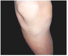
사진 260. 무릎 관절
Copyright ⓒ 2011 John Sangwon Lee, M.D., FAAP
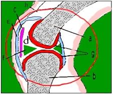
그림 263. 무릎 관절의 구조
a-대퇴골, b-경골, c-슬개골, d-관절낭, e-활막, f-메니쿠스, g-연골, h-인대
Copyright ⓒ 2011 John Sangwon Lee, M.D., FAAP
-
무릎관절이 외상으로 관절을 형성하고 있는 대퇴골, 경골, 비골, 슬개골 등이 골절될 수 있고 탈골될 수 있다.
-
슬개골 탈골은 사춘기 아이들에게 잘 생길 수 있고 남아들보다 여아들에게 더 잘 생긴다.
-
걸을 때 무릎관절 이하 아랫다리가 외반될 수 있다.
-
무릎이 아프고 슬개골 부위를 만지면 뽀각뽀각 소리가 나고 압통이 생길 수 있다.
-
탈골 전 슬개골이 자연적으로 제자리로 돌아갈 수 있다.
-
슬개골이 갑자기 탈골될 때는 손으로 만져 복구 치료를 할 수 있다.
-
의사의 지시에 따라 약 3주 동안 무릎을 더 이상 움직이지 말고 무릎을 쓰지 않는 부동치료를 한다.
-
재발되면 수술로 고정치료 한다.
-
이상 설명한 무릎 관절에 생긴 외상에 관해서는 이 정도로 설명하고 다음에는 주로 무릎 관절을 형성하고 있는 뼈에 생긴 골절에 대해서 설명한다.
무릎 관절이 다칠 때의 증상 징후
-
무릎 관절을 형성하고 있는 연조직이나 여러 종류의 뼈들 중(그림 7.53, 263 참조) 어느 뼈가 어느 정도 골절되었는지에 따라 증상 징후가 다르다.
-
무릎 관절 속에 있는 뼈가 골절되면 무릎이 붓고, 아프며, 골절된 부위를 손으로 누르거나 능동적으로나 수동적으로 움직이면 상당히 아픈 것이 보통이다.
-
그 때문에 골절된 무릎을 능동적으로 수동적으로 굽혔다 폈다 하기가 대단히 곤란하다.
-
어떤 때는 골절된 뼈끝이 서로 어긋날 수 있고, 골절된 뼈끝이 서로 떨어지고 분리될 수 있다.
-
골절된 뼈의 양쪽 끝 사이를 손으로 만져보면 그 사이 속으로 손가락 하나가 들어갈 수 있을 정도로 골절된 양 뼈끝 사이에 간격이 넓게 벌어질 수 있다.
-
심한 무릎 관절을 형성하고 있는 뼈가 골절되어 있을 때 골절된 뼈끝이 피부층을 꿰뚫고 피부층 밖으로 삐져나올 수도 있다.
무릎 관절 속 뼈가 골절될 때의 진단
-
병력, 증상 징후와 진찰소견 등을 종합해서 무릎 관절의 뼈가 골절되었다고 의심되면 무릎 관절 X선 사진 검사 등으로 진단한다.
무릎 관절 속 뼈가 골절될 때의 치료
-
무릎 관절을 심하게 다쳐 무릎 관절에 있는 뼈가 골절되었다고 생각하면 의사의 지시가 없이 가능한 한 그 무릎을 더 이상 움직이지 말아야 한다.
-
가능한 한 다친 무릎을 처음 목격한 상태 그대로 두고 의료구급대나 병원 응급실이나 의사에게 긴급 전화 진료 상담을 해서 그들의 지시에 따라 현장에서 치료를 시작한다.
-
침착하게 환아를 안정시킨다.
-
의사나 구급대원의 지시에 따라 구급차나 다른 적절한 교통수단을 이용해 병원 응급실로 급히 데리고 간다.
-
무릎 관절 속에 있는 뼈가 골절되었다고 의심되면 그 다리 전체에 혈액순환이 잘되는지 체크해 보기 위해서 다친 무릎 아래, 위의 피부의 색을 관찰해 의사에게 보고하면서 의사의 지시에 따라 응급처치 한다.
-
발등에 있는 발등 동맥에서 맥을 짚어보고 그쪽 다리의 혈액순환이 정상인지 확인해서 의사에 보고하면서 병원으로 후송한다.
-
구급대원이나 의사가 현장으로 올 수 없을 때는 상황에 따라 발목 관절뼈 골절을 치료할 때와 같이 처음 목격했을 때의 상태의 자세 그대로 무릎 관절을 유지하면서 골절된 무릎 관절 속의 뼈와 그 관절의 아래 위 부위, 그쪽 아랫다리 전체와 허벅다리 전체에 부목을 대어 골절된 무릎을 고정시킨 후에 병원 응급실로 데리고 갈 수 있다.
-
이 처치도 될 수 있으면 의사의 지시에 따라서 한다.
-
만약에 골절된 무릎 관절뼈의 아랫다리가 창백하거나 파랗거나 감각이 없을 때는 조금도 지체하지 말고 병원 응급실이나 의사에게 긴급으로 전화해 그들의 도움을 응급하게 받으면서 구급차로나 다른 적절한 교통수단 방법으로 병원 응급실로 급히 데리고 가는 것이 이상적 응급처치이다.
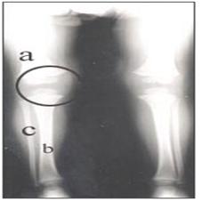
사진 264. ◯로 표시한 부위에 무릎 관절
a-대퇴골, b-경골, c-비골
Copyright ⓒ 2011 John Sangwon Lee, M.D., FAAP

사진 265. ◯로 표시한 부위에 무릎 관절
a-대퇴골, b-경골, c-비골, d-무릎 관절, e ◯내–경골 골절
Copyright ⓒ 2011 John Sangwon Lee, M.D., FAAP
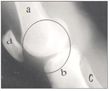
사진 266.정상 무릎 관절의 측면 X선 사진
◯로 표시한 부위에 무릎관절이 있다.
a-대퇴골, b-경골, c-비골, d-슬개골
Copyright ⓒ 2011 John Sangwon Lee, M.D., FAAP
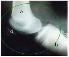
사진 267. 무릎 관절의 측면 X선 사진
◯로 표시한 부위에 무릎 관절
a-대퇴골, b-경골, c-비골, d-슬개골
경골 두부와 비골 두부가 탈구됐다.
Copyright ⓒ 2011 John Sangwon Lee, M.D., FAAP
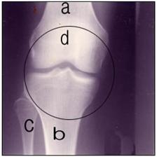
사진 268. 정상 무릎 관절의 전후면 X선 사진
◯내 무릎 관절
a-대퇴골, b-경골, c-비골, d-슬개골
Copyright ⓒ 2011 John Sangwon Lee, M.D.. FAAP
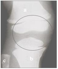
사진 269. 정상 무릎 관절의 전후면 X선 사진
◌내에 무릎 관절
a-대퇴골, b-경골, c-비골
Copyright ⓒ 2011 John Sangwon Lee, M.D., FAAP
Patellar trauma and patellar dislocation 슬개골 외상과 슬개골 탈골
Photo 260. Knee joint Copyright ⓒ 2011 John Sangwon Lee, M.D., FAAP
Figure 263. Structure of the knee joint a-femur, b-tibia, c-patella, d-capsule, e-synovial membrane, f-menicus, g-cartilage, h-ligament Copyright ⓒ 2011 John Sangwon Lee, M.D., FAAP
• As a result of trauma to the knee joint, the femur, tibia, fibula, and patella can be fractured and dislocated.
• Patellar dislocation is more likely to occur in adolescents and is more common in girls than boys.
• When walking, the lower leg below the knee joint may be valgus.
• If your knee hurts and you touch the patella area, it may make a clicking sound and tenderness.
• The kneecap can naturally return to its original position before dislocation.
• When the patella is suddenly dislocated, you can touch it with your hands for recovery treatment.
• According to the doctor’s instructions, do not move the knee any longer and perform knee immobilization treatment for about 3 weeks.
• In case of recurrence, fixed treatment is performed by surgery.
• The trauma to the knee joint described above will be explained to this extent, and then the fractures in the bones that mainly form the knee joint will be explained.
Symptoms Signs When Your Knee Joint Injures
• Among the soft tissues forming the knee joint or various types of bones (see figs. 7.53 and 263), the symptoms differ depending on which bone is fractured and to what extent.
• When a bone in the knee joint is fractured, the knee is swollen and painful, and it is usually quite painful when you press the fractured area with your hand or move it actively or passively.
• As a result, it is very difficult to actively and passively bend and straighten the fractured knee.
• At times, the fractured tip may be displaced, and the fractured tip may fall apart and separate.
• If you touch the ends of the fractured bones with your hands, the gap between the ends of the fractured bones can be wide enough that one finger can fit into the gap.
• When the bones forming the severe knee joint are fractured, the fractured bone tip may penetrate the skin layer and protrude out of the skin layer.
Diagnosis of fractures of bones in the knee joint
• If it is suspected that the bone of the knee joint is fractured based on the medical history, symptom signs, and examination findings, it is diagnosed with an X-ray examination of the knee joint.
Treatment for fractured bones in the knee joint
• If you have seriously injured your knee joint and think you have fractured a bone in the knee joint, you should stop moving the knee as much as possible without your doctor’s instructions.
• If possible, leave the injured knee as it was at the first sighting, and make an emergency phone consultation to a paramedic, hospital emergency room, or doctor, and follow their instructions to begin treatment on the spot.
• Calmly stabilize the child.
• As directed by a doctor or paramedic, take them quickly to the hospital emergency room by ambulance or other suitable means of transportation.
• If you suspect that a bone in the knee joint is fractured, observe the color of the skin below and above the injured knee to check whether blood circulation is good throughout the leg, report it to the doctor, and take first aid according to the doctor’s instructions.
• Take a pulse from the dorsal artery on the dorsum of the foot, check that blood circulation in that leg is normal, report it to the doctor, and transport him to the hospital.
• When paramedics or doctors cannot come to the scene, depending on the circumstances, such as when treating an ankle joint fracture, the knee joint is maintained in the same posture as when it was first observed, and the bones in the fractured knee joint and the lower and upper joints After the fractured knee is immobilized by splinting the area, the entire lower leg and the entire thigh, the patient can be taken to the hospital emergency room.
• If possible, this treatment should be done as directed by your doctor.
• If the lower leg of the fractured knee joint is pale, blue, or numb, do not delay at all and immediately call the hospital emergency room or doctor for immediate assistance, and use an ambulance or other suitable means of transportation to get to the hospital emergency room. Taking them quickly is the ideal first aid.
Picture 264. Knee joint in the area marked with ◯ a – femur, b – tibia, c – fibula Copyright ⓒ 2011 John Sangwon Lee, M.D., FAAP
Picture 265. Knee joint in the area marked with ◯ a-femur, b-tibia, c-fibula, d-knee joint, e ◯internal-tibia fracture Copyright ⓒ 2011 John Sangwon Lee, M.D., FAAP
Picture 266. Lateral X-ray of a normal knee joint There is a knee joint in the area marked with ◯. a-femur, b-tibia, c-fibula, d-patella Copyright ⓒ 2011 John Sangwon Lee, M.D., FAAP
Picture 267. Lateral X-ray of the knee joint in the area marked with ◯ a-femur, b-tibia, c-fibula, d-patella The tibialis head and fibula head were dislocated. Copyright ⓒ 2011 John Sangwon Lee, M.D., FAAP
Picture 268. Anterior and posterior X-ray of a normal knee joint ◯My knee joint a-femur, b-tibia, c-fibula, d-patella Copyright ⓒ 2011 John Sangwon Lee, M.D., FAAP
Picture 269. Anterior and posterior X-ray of a normal knee joint ◌Inside the knee joint a – femur, b – tibia, c – fibula Copyright ⓒ 2011 John Sangwon Lee, M.D., FAAP
출처 및 참조 문헌 Sources and references
- NelsonTextbook of Pediatrics 22ND Ed
- The Harriet Lane Handbook 22ND Ed
- Growth and development of the children
- Red Book 32nd Ed 2021-2024
- Neonatal Resuscitation, American Academy Pediatrics
-
Childhood Emergencies in the Office, Hospital and Community, American Academy of Pediatrics
-
Emergency Medical Service for Children, By Ross Lab. May 1989. p.10
-
Emergency care, Harvey grant, and Robert Murray
-
Emergency Care Transportation of Sick and Injured American Academy of Orthopaedic Surgeons
-
Emergency Pediatrics A Guide to Ambulatory Care, Roger M. Barkin, Peter Rosen
-
Immediate care of the acutely ill and injured, Hugh E. Stephenson, Jr
-
The Critically Ill Child, Diagnosis and Management, Edited by Clement A. Smith
-
Emergency Medical Services for Children: The Role of the Primary Care Provider, America Academy of Pediatrics
-
Quick Reference To Pediatric Emergencies, Delmer J. Pascoe, M.D., Moses Grossman, M.D. with 26 contributors
-
Manual of Emergency Care 응급환자관리 정담미디어
-
소아가정간호백과–부모도 반의사가 되어야 한다, 이상원
-
Neonatal Resuscitation American heart Association
-
Neonatology Jeffrey J.Pomerance, C. Joan Richardson
-
Pediatric Resuscitation Pediatric Clinics of North America, Stephen M. Schexnayder, M.D.
-
Pediatric Critical Care, Pediatric Clinics of North America, James P. Orlowski, M.D.
-
Preparation for Birth. Beverly Savage and Dianna Smith
-
Nelson Textbook of Pediatrics 14th ed. Beherman,
-
The Johns Hopkins Hospital, The Harriet Lane Handbook, 18th edition
-
Red book 29th edition 2012
-
Nelson Text Book of Pediatrics 19th Edition
-
Infectious disease of children, Saul Krugman, Samuel L Katz, Ann A. Gershon, Catherine Wilfert
-
The Harriet Lane Handbook 19th Edition
-
소아과학 안효섭 외 대한교과서
-
제1권 소아청소년 응급의료 참조문헌과 출처
-
Other