- 손바닥뼈는 수근골(팔목 뼈에 연결된 뼈)과 중수골(손 중간에 있는 뼈)로 형성되고 손가락뼈는 지절골(손가락뼈)로 형성된다.
- 이 손바닥뼈와 손가락뼈들 중 어느 뼈도 골절될 수 있다.
-
손뼈의 골단, 골간 등 어느 부위가 골절될 수 있다.
-
운동, 장난, 또는 안전사고 등으로 손에 있는 뼈와 뼈 사이에 있는 관절 또는 손가락 마디 관절(수지 관절)이 삘 수 있고, 또 삐기도 하고 골절이 동시 생길 수 있다.
-
특히 아이들이 육체적 운동을 하다가 손이나 손가락, 또는 손목에 있는 근대나 건이 손상 될 수 있고, 손에 있는 뼈에 골절이 생길 수 있고, 관절이 삐는 등 여러 가지 외상을 입을 수 있다.
손바닥 뼈골절과 손가락뼈 골절의 증상 징후
-
손바닥뼈의 골절의 정도와 골절된 부위 등에 따라 증상 징후가 다르다.
-
일반적으로 골절된 부위가 붓고 만지면 아프며 골절이 되고 외상 입은 손 부위를 정상적으로 움직일 수 없다.
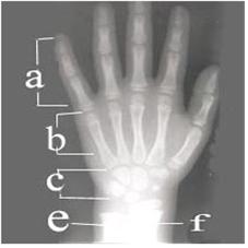
사진 124. 손바닥뼈와 손가락뼈 X-선 사진
손뼈를 수근골 (Carpus), 중수골 (Metacarpus), 지골(Phalages)로 3분 한다.
좌우 손의 X-선 사진에서 보는 바와 같이 각 손뼈의 크기와 모양이 나이에 따라 다르다.
특히 아이의 나이에 따라 수근골의 크기와 수가 다르다.
수근골의 X-선 사진은 소아 골연령(Bone age)을 알아보는 데 사용 한다.
a-지골, b-중수골, c-수근골, e-척골, f-요골
Copyright ⓒ 2013 John Sangwon Lee, M.D., FAAP
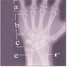
사진 125. 손바닥뼈와 손가락의 X-선 사진
좌우 손 X-선 사진에서 보는 바와 같이 각 뼈의 크기와 모양이 나이에 따라 다르다. 특히 아이의 나이에 따라 수근골의 크기와 수가 다르다.
a-지골, b-중수골, c-수근골, e-척골, f-요골
Copyright ⓒ 2013 John Sangwon Lee, M.D., FAAP
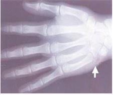
사진 127. 10세 된 학령기 아이의 수근골 골절과 정상 손가락뼈
↑로 가리킨 부위의 수근골이 골절됐다.
Copyright ⓒ 2012 John Sangwon Lee, M.D., FAAP
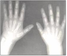
사진 126. 1세 된 유아의 정상 양 손바닥뼈와 손가락뼈
Copyright ⓒ 2012 John Sangwon Lee, M.D., FAAP
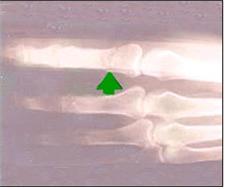
사진 129. 골절된 손가락 뼈(↑로 표시된 부분)
Copyright ⓒ 2013 John Sangwon Lee, M.D., FAAP
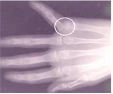
사진 128. 손가락뼈와 중수골 관절이 탈구되고 골절도 됐다(◯내)
Copyright ⓒ 2013 John Sangwon Lee, M.D., FAAP
손바닥 뼈골절과 손가락뼈의 골절의 진단
-
병력, 증상 징후와 진찰소견 등을 종합해서 손바닥뼈(수근골와 중수골)나
-
손가락뼈(수지골/ 지골)가 골절됐다고 의심되면 손바닥 뼈나 손가락 뼈 X-선 사진 검사로 쉽게 진단한다.
-
손바닥뼈나 손가락뼈 골절이 있을 때의 증상 징후가 있는데도 X-선 검사에는 골절이 보이지 않을 수 있다.
-
이런 때는 골절이 있다고 추정 진단을 하고 손바닥뼈 골절이나 손가락뼈 골절 치료를 하는 것과 같이 우선 적절히 치료하고 첫 X-선 검사를 한 후 7-10일 후에 추적 X선 검사를 해서 손뼈 골절을 확진할 때도 있다.
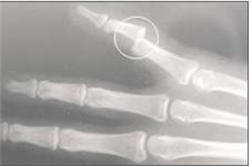
사진131. 탈구도 되고 골절된 손가락뼈 (○ 내)
Copyright ⓒ 2013 John Sangwon Lee, M.D., FAAP
손바닥 뼈골절과 손가락 뼈 골절의 치료
-
손바닥 뼈 골절이나 손가락뼈의 골절의 정도에 따라 치료가 다르다.
-
삐거나 골절된 손바닥 뼈나 손가락 뼈, 또는 손가락 관절을 적기에 적절히 치료하지 않으면 손바닥이나 손가락에 기형이 생길 수 있고 기능 장애가 생길 수 있다.
-
그런 기형이 영구적으로 지속 될 수 있다.
-
손바닥이나 손가락의 기능이 영구적으로 상실될 수 있다.
-
이런 이유로 손뼈나 손가락뼈가 골절되거나 손에 외상이 생기면 손뼈 골절 등을 특별히 전문하는 정형외과 전문의의 적절한 치료를 받는 것이 상당히 중요하다.
-
손에 외상을 입으면 가능하면 우선 환아를 안정시키고 안전한 곳으로 옮긴다.
-
손가락뼈나 손바닥 뼈가 골절되었다고 의심되면 의료구급대원이나 의사나 병원 응급실에 긴급 전화 진료 상담을 해서 그들의 지시에 따라 현장에서 치료를 시작한다.
-
손바닥뼈 골절의 정도에 따라 적절한 교통수단을 이용해서 종합 병원 응급실로 데리고 간다.
-
손바닥 뼈 골절이 생겼다고 의심하면 손가락과 그 주위의 정상 손가락, 손, 팔목, 아래팔까지 부목을 대어 응급처치를 할 수 있다.
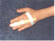
사진 132. 손가락뼈나 손바닥뼈가 골절됐다고 의심하면 그 부위를 누르지도 밀지도 당기거나 틀지도 말고 있는 상태에서 부목을 대고 병원으로 갈 수 있다.
Copyright ⓒ 2013 John Sangwon Lee, M.D., FAAP
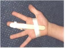
사진 133. 손가락뼈나 손바닥뼈가 골절됐다고 의심되면 그 부위를 누르지도 밀지도 당기거나 틀지도 말고 있는 그 상태에서 부목을 대고 병원으로 갈 수 있다.
Copyright ⓒ 2013 John Sangwon Lee, M.D., FAAP
-
부목을 대기 전과 대는 중 댄 후에 골절된 손가락과 손의 피부색, 그쪽 손톱 밑 혈색, 손이나 손가락에 분포된 말초신경 기능이 정상인지 체크해 본다.
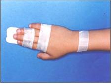
사진 134. 손바닥뼈나 손가락뼈가 골절됐다고 의심되면 있는 그 상태에서 부목을 대고 병원으로 가든지 의사의 지시에 따라 응급 치료한다.
Copyright ⓒ 2013 John Sangwon Lee, M.D., FAAP
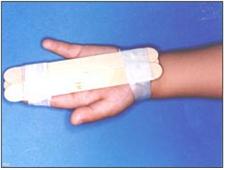
사진 135. 손뼈나 손가락뼈에 골절됐다고 의심되면 있는 그 상태에서 부목을 대고 병원으로 가든지 의사의 지시에 따라 응급 치료한다.
Copyright ⓒ 2013 John Sangwon Lee, M.D., FAAP
-
손이나 손가락이 파랗거나 거기에 아무 감각이 없거나 자유자재로 움직일 수 없으면 현장에서 단골 소아청소년과 의사나 병원 응급실에 긴급 문의 진료 상담을 해 그들의 지시대로 응급처치를 한다.
-
그렇지 않으면 병원 응급실로 급히 데리고 간다.
-
손바닥뼈나 손가락뼈가 골절되었을 때 적절한 시기에 적절히 치료하지 않으면 손이나 손가락의 기능 장애가 생겨 평생 불구자가 될 수 있다고 이미 언급했다.
-
특히, 소아 지골 골절을 적절히 치료하지 않으면 회전변형이 생길 수 있다.
-
금만 간 지골 골절은 2~3주 동안 부목으로 치료하고 3~4주 동안 골절된 지골의 바로 옆에 있는 다음 손가락을 함께 테이프 고정으로 치료한다.
-
중수골이 골절됐을 때는 중수골이 골절된 정도에 따라 캐스트 고정 치료를 하거나 부목으로 치료하든지, 또는 수술로 치료 할 수 있다.
-
따라서 손바닥뼈나 손가락뼈가 골절되었거나, 마디가 탈구되었거나, 삐었을 때는 일반 정형외과 전문의나 손뼈 골절 전문 정형외과 전문의의 치료를 받는다.
|
다음은“새끼 손가락 좌상”에 관한 인터넷 소아청소년 건강 상담 질의응답 입니다. |
Q&A. 새끼 손가락 좌상에 대한 문의,
Q.
- 생후 8개월 된 여아입니다.
- 방문 틈에 세끼손가락이 껴있는 상태에서 오빠가 문이 닫힐 정도로 세게 닫았습니다.
- 동네 의원에서 엑스레이를 찍었는데 애기가 가만히 있지를 않아 뼈 상태는 확실하지 않다고 하고 괜찮을 거라고 합니다.
- 손가락 마디마디가 부어있고 멍이 들어있으며 손끝은 핏기가 있습니다.
- 성장판이 손상되면 손가락도 자라지 않는다고 하는데 어떻게 하면 확인을 할 수 있는지요. 혹시 뼈에 이상이 있을 수 있나요. 꼭 답변 부탁드립니다.
A.
- 지원님
- 안녕하세요. 질문해 주셔서 감사합니다. 좋은 질문입니다.
- 자녀의 나이, 성별, 과거 병력, 가족 병력, 진찰소견, 임상검사 등의 정보를 많이 알수록 답을 드리는데 도움이 됩니다. 주신 정보를 토대로 해서 답변을 드리겠습니다.
- 자세한 정보를 주셔서 감사합니다.
- 8개월 된 영아들의 신체의 어떤 부위 X-사진을 찍는 것은 때로는 여간 힘든 일이 아닙니다.
- 아기가 낮잠을 자는 동안에 X-선 사진을 다시 한 번 찍어보는 것이 좋을 것입니다.
- 드물게는 정신 안정제로 안정시키고 뼈 X-사진 검사를 할 때도 있습니다.
- 지금은 골절이 있든지 없든지 2주정도 부목을 손가락과 손바닥에 대 고정 치료를 하는 것도 좋을 것입니다.
- 요즘 정형외과에 소아 정형외과도 있고 성인 정형외과도 있습니다.
- 소아 정형외과 전문의들 중 소아 손 정형외과를 특별히 더 전문하는 정형외과 전문의도 있습니다.
- 필요 하면 단골 소아청소년과 의사에 부탁해 손 정형외과 전문의의 자문을 받으셔요.
- 손뼈와 손가락뼈의 골절, 골절, 등을 참조하시기 바랍니다. 질문이 더 있으면 다시 연락해 주시기 바랍니다. 감사합니다. 이상원 드림
Fracture of hand bone and finger bone(Hand and finger fractures) 손뼈 골절과 손가락뼈 골절
• The palmar bone is formed from the carpal (a bone connected to the wrist bone) and metacarpal (a bone in the middle of the hand), and the finger bone is formed from the phalanges (finger bones).
• Any of these metacarpals and finger bones can be fractured.
• Any part of the hand bone, such as the epiphysis or the diaphysis, may be fractured.
• The joint between the bones in the hand or the knuckle joint (digital joint) may be sprained, or sprained and fractured at the same time due to exercise, play, or a safety accident.
• Children, especially during physical exercise, can damage the hamstrings or tendons in their hands, fingers, or wrists, fracture bones in their hands, and sprain their joints.
Symptoms, signs of Palmar Fractures and Finger Bone Fractures
• Symptoms and signs vary depending on the degree of fracture of the metacarpal and the site of the fracture.
• In general, the fractured area is swollen and painful to the touch, and the fractured area is unable to move normally in the injured hand area.
Picture 124. X-ray of the metacarpal and finger bones The hand bones are divided into three segments: carpus, metacarpus, and phalanges. As shown in the X-ray images of the left and right hands, the size and shape of each hand bone differs according to age. In particular, the size and number of carpal bones vary according to the age of the child. X-rays of the carpal bones are used to determine the bone age of children. a-phalange, b-metacarpal, c-carpal, e-ulnar, f-radius Copyright ⓒ 2013 John Sangwon Lee, M.D., FAAP
Picture 125. X-ray picture of the metacarpal and fingers As you can see in the left and right hand X-rays, the size and shape of each bone varies with age. In particular, the size and number of carpal bones vary according to the age of the child. a-phalange, b-metacarpal, c-carpal, e-ulnar, f-radius Copyright ⓒ 2013 John Sangwon Lee, M.D., FAAP
Picture 127. Carpal fracture and normal finger bones in a 10-year-old school-age child The carpal bone in the area indicated by ↑ was fractured. Copyright ⓒ 2012 John Sangwon Lee, M.D., FAAP
Photo 126. Normal both metacarpals and finger bones of a 1-year-old infant Copyright ⓒ 2012 John Sangwon Lee, M.D., FAAP
Picture 129. Fractured finger bones (marked with ↑) Copyright ⓒ 2013 John Sangwon Lee, M.D., FAAP
Picture 128. The finger and metacarpal joints were dislocated and fractured (within ◯) Copyright ⓒ 2013 John Sangwon Lee, M.D., FAAP
Diagnosis of Fractures of the Palmare and Fractures of the Finger Bone
• Combining the medical history, symptom signs, and examination findings, the palmar bone (carpal and metacarpal)
• If a finger bone (digital bone/phalanx) is suspected to be fractured, it is easily diagnosed with an X-ray examination of the palmar bone or finger bone.
• X-rays may not show a fracture even when there are signs of symptoms of a fracture of the metatarsal or finger bones.
• In this case, it is possible to make a presumptive diagnosis of a fracture, treat it appropriately, such as treating a metacarpal fracture or a finger bone fracture, and perform a follow-up X-ray 7-10 days after the first X-ray examination to confirm the hand fracture sometimes
Picture 131. Dislocated and fractured finger bones (within ○) Copyright ⓒ 2013 John Sangwon Lee, M.D., FAAP
Treatment of palmar fractures and finger bone fractures
• Treatment varies depending on the severity of the fracture of the palmar bone or the fracture of the finger bones.
• If the sprained or fractured metacarpal, finger bones, or knuckles are not treated in a timely and appropriate manner, the palm or fingers can become deformed and dysfunctional.
• Such malformations can be permanent. • Permanent loss of function of the palm or fingers.
• For this reason, it is very important to receive appropriate treatment from an orthopedic surgeon who specializes in hand bone fractures, etc., if the hand bones or finger bones are fractured or there is a trauma to the hand.
• In case of hand trauma, first stabilize the child and move him/her to a safe place if possible.
• If you suspect that you have fractured a finger or palmar bone, consult a paramedic, doctor, or emergency phone call emergency room at the hospital, and follow their instructions to start treatment on the spot.
• Depending on the severity of the metacarpal fracture, take them to the emergency room of a general hospital by means of appropriate transportation.
• If you suspect a metacarpal fracture has occurred, you can provide first aid by splinting the finger and the surrounding normal fingers, hand, wrist, and forearm.
Photo 132. If you suspect that you have fractured a finger or metacarpal, you can apply a splint and go to the hospital without pressing, pushing, pulling, or twisting the area. Copyright ⓒ 2013 John Sangwon Lee, M.D., FAAP
Photo 133. If you suspect that you have fractured a finger or metacarpal, you can apply a splint and go to the hospital without pressing, pushing, pulling, or twisting the area. Copyright ⓒ 2013 John Sangwon Lee, M.D., FAAP
• Before and after applying the splint, check whether the skin color of the fractured finger and hand, the color under the fingernail, and the peripheral nerve function distributed in the hand or finger are normal.
Photo 134. If you suspect that you have fractured the metacarpal or finger bones, apply a splint while still present and go to the hospital or receive emergency treatment according to the doctor’s instructions. Copyright ⓒ 2013 John Sangwon Lee, M.D., FAAP
Photo 135. If you suspect a fracture of the hand or finger bones, apply a splint while still present and go to the hospital or receive emergency treatment according to the doctor’s instructions. Copyright ⓒ 2013 John Sangwon Lee, M.D., FAAP
• If your hands or fingers are blue, there is no sensation there, or you cannot move freely, consult with your regular pediatrician or hospital emergency room on the spot and provide first aid according to their instructions.
• Otherwise, take him to the hospital emergency room.
• We have already mentioned that fractures of the metacarpal or finger bones can lead to life-long disability if not properly treated at the right time, resulting in hand or finger dysfunction.
• Rotational deformation can occur, especially if the fracture of the phalanx in children is not properly treated.
• Cracked phalanx fractures are treated with a splint for 2-3 weeks and the next finger next to the fractured phalanx is treated with tape fixation for 3-4 weeks.
• When the metacarpal is fractured, it can be treated with cast fixation, splints, or surgery depending on the degree of fracture of the metacarpal.
• Therefore, when a metacarpal or finger bone is fractured, a joint is dislocated, or is sprained, seek treatment from a general orthopedic specialist or an orthopedic specialist specializing in hand bone fractures.
The following is a Q&A on the Internet Pediatric Health Counseling regarding “Strained Little Finger”.
Q&A. Inquiries about a left little finger,
Q.
• This is an 8 month old girl.
• Her brother slammed the door shut with three fingers stuck in the gap between the door.
• She had X-rays taken at the local doctor and she said the baby was not still and her bones were not sure and she said she would be fine. • Her knuckles are swollen and bruised, and her fingertips are bloody.
• It is said that if the growth plate is damaged, the fingers do not grow. How can I check it? Could there be something wrong with the bones? Please do reply.
A.
• Support • Good morning. Thanks for asking. That’s a good question.
• The more information you have, such as your child’s age, gender, past medical history, family history, examination findings, and clinical tests, it will help you to give an answer. We will give you an answer based on the information you provided.
• Thanks for the detailed information.
• Taking X-photographs of any part of the body of 8 month old infants is sometimes not a daunting task.
• It might be a good idea to take another x-ray while your baby is napping.
• Rarely, he is stabilized with a sedative and a bone x-graph is done.
• Now, whether or not there is a fracture, it would be good to fix the splint on the finger and palm for about 2 weeks.
• Orthopedics these days include pediatric orthopedics and adult orthopedics.
• Some pediatric orthopedic specialists specialize in pediatric hand orthopedic surgery specifically.
• If necessary, ask your regular pediatrician for advice from a hand orthopedic surgeon.
• See Fractures, Fractures, etc. of the hand and finger bones. If you have any more questions, please contact us again. Thank you. Lee Sang-won. MD.
출처 및 참조 문헌 Sources and references
- NelsonTextbook of Pediatrics 22ND Ed
- The Harriet Lane Handbook 22ND Ed
- Growth and development of the children
- Red Book 32nd Ed 2021-2024
- Neonatal Resuscitation, American Academy Pediatrics
- www.drleepediatrics.com 제16권 소아청소년 정형외과 질환
- www.drleepediatrics.com제8권 소아청소년 호흡기 질환
- www.drleepediatrics.com제9권 소아청소년 소화기 질환
- www.drleepediatrics.com제10권. 소아청소년 신장 비뇨 생식기 질환
- www.drleepediatrics.com제11권. 소아청소년 심장 혈관계 질환
- www.drleepediatrics.com제12권. 소아청소년 신경 정신 질환, 행동 수면 문제
- www.drleepediatrics.com제13권. 소아청소년 혈액, 림프, 종양 질환
- www.drleepediatrics.com제14권. 소아청소년 내분비, 유전, 염색체, 대사, 희귀병
- www.drleepediatrics.com제15권. 소아청소년 알레르기, 자가 면역질환
- Red book 29th-31st edition 2021
- Nelson Text Book of Pediatrics 19th — 21st Edition
- The Johns Hopkins Hospital, The Harriet Lane Handbook, 22nd edition
-
Quick Reference to Pediatric Emergencies, Delmer J. Pascoe, M.D., p.160-161
-
Emergency Pediatrics, A guide to ambulatory care, 5th edi. Roger M. Barkin, Peter Rosen, p.536-537
-
Emergency care and transportation of the sick and injured,3rd edition, American Academy of orthopedic surgeons. p.129, 275-276
-
Nelson text book, 14th edition, p.80, 1722, 1723-1728
-
Childhood Emergencies in the Office, Hospital and Community, American Academy of Pediatrics
-
Emergency Medical Service for Children, By Ross Lab. May 1989. p.10
-
Emergency care, harvey grant and robert murray
-
Emergency Care Transportation of Sick and Injured American Academy of Orthopaedic Surgeons
-
Emergency Pediatrics A Guide to Ambulatory Care, Roger M. Barkin, Peter Rosen
-
Immediate care of the acutely ill and injured, Hugh E. Stephenson, Jr
-
The Critically Ill Child, Diagnosis and Management, Edited by Clement A. Smith
-
Emergency Medical Services for Children: The Role of the Primary Care Provider, America Academy of Pediatrics
-
Quick Reference To Pediatric Emergencies , Delmer J. Pascoe, M.D., Moses Grossman, M.D. with 26 contributors
-
Manual of Emergency Care
-
응급환자관리 정담미디어
-
소아가정간호백과–부모도 반의사가 되어야 한다, 이상원
-
Neonatal Resuscitation American heart Association
-
Neonatology Jeffrey J.Pomerance, C. Joan Richardson
-
Pediatric Resuscitation Pediatric Clinics of North America, Stephen M. Schexnayder, M.D.
-
Pediatric Critical Care, Pediatric Clinics of North America, James P. Orlowski, M.D.
-
Preparation for Birth. Beverly Savage and Dianna Smith
- Infectious disease of children, Saul Krugman, Samuel L Katz, Ann A. Gerhon, Catherine Wilfert
-
The Harriet Lane Handbook 19th Edition
-
소아과학 대한교과서
-
제1권 소아청소년 응급의료 참조문헌과 출처
-
Other
Copyright ⓒ 2015 John Sangwon Lee, MD., FAAP
“부모도 반의사가 되어야 한다”-내용은 여러분들의 의사로부터 얻은 정보와 진료를 대신할 수 없습니다.
“The information contained in this publication should not be used as a substitute for the medical care and advice of your doctor. There may be variations in treatment that your doctor may recommend based on individual facts and circumstances. “Parental education is the best medicine.”