소화관 내 이물 Foreign bodies in digestive tracts
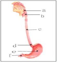
그림 202. 소화관 내 이물
a-인두강 내 이물, b, c, d-식도관 내 이물, e-위 내 이물, f-소장 내 이물
출처-Used with permission from Galaxo Wellcome
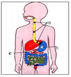
그림 203. 소화관 내 이물
a, b, c-식도관 내 이물, d-위 내 이물, e-소장관 내 이물
Copyright ⓒ 2011 John Sangwon Lee, MD., FAAP
소화관 내 이물
- 인두 강 내 이물, 식도관 내 이물, 위 내 이물, 소대장관 내 이물을 통털어 소화관 내 이물이라고 한다.
- 통계에 의하면, 소아청소년들의 소화관 내 이물과 기도 내 이물의 33%는 기관 내 이물과 기관지 내 이물이고, 56%는 식도관 내 이물과 그 외 소화관 내 이물이었다.
- 인두강 내 이물, 식도관 내 이물, 위 내 이물(위 속 이물), 소장관 내 이물, 대장관 내 이물 등 소화관 내 이물의 대부분은 동전, 단추, 작은 금속 덩어리, 안전핀 등이었고,
- 기도 내 이물의 대부분은 땅콩, 콩, 팝콘, 핀, 나사, 철사, 못, 연필 지우개 등이었다. 카메라, 계산기, 시계 등에 들어 있는 디스크 배터리(디스크 전지)를 삼켜 식도관 내에 걸쳐 식도관 내 이물이 될 수 있고 위 내 또는 소장관 내에 있어 위장관 내 이물이 될 수 있다.
- 기도 내 이물의 대부분은 5세 이하 영유아들에게 가장 많이 생겼고, 소화관 내 이물의 대부분은 6개월∼3세 사이의 영유아들에게 가장 많이 생겼다고 한다.
- 영유아들이 이물을 잘못 삼켜서 기도 내나 소화관 내로 들어가지 않도록 각별히 유의하고 미리부터 예방해야 한다.
- 입 안, 인두, 식도, 위, 십이지장, 소장, 대장 등은 소화기(소화기 계통/ 소화 계통)에 속하고 거기에 생긴 이물을 소화관 내 이물 또는 소화관 이물이라고 한다.
표 8. 소아들의 소화관 내 이물
| 소화관 내 이물의 종류 | 소화관 내 이물 |
| 음식물 | 복숭아 씨 등 과일의 씨, 고기 뼈 조각, 생선 가시나 뼈 등 |
| 나무 | 나무 조각, 이쑤시개, 나무 장난감 등의 부속물 |
| 전지 | 카메라 전지, 계산기나 시계 등의 디스크 전지(디스크 배테리) 등 |
| 보석류 | 유리조각, 귀걸이, 반지 등 작은 보석, 공기, 구슬 등 |
| 화장품 | 펜캡, 연필, 고무 지우게, 크레용, 종이클립 등 |
| 장남감 | 구슬, 공기 펜캡 등 |
| 그 외 | 동전, 바늘, 핀, 위 내 털뭉치 등 |
소화관 내 이물(인두 강 이물, 식도관 내 이물, 위 내 이물, 장관 내 이물/위장관 내 이물)의 증
상 징후
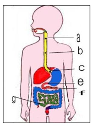
그림 204. 식도관 내나 위장관 내 이물은 그림 a, b, c, e, f, g 생리적으로 협소한 식도관 내와 위장관 내 부분에 걸려 있을 수 있다.
a-윤상 인두,
b-대동맥 아취,
c-분문,
e-유문,
f-트라이즈 인대,
g-회맹장.
CopyrightⒸ 2011 John Sangwon Lee,MD., FAAP
-
영유아들은 호기심이 많고 사리판단이 미숙하기 때문에 손에 닥치는 대로 입안에 넣을 수 있을 정도로 작은 물체는 거의 무엇이든 입안에 넣고 빨고 노는 버릇이 있다.
-
입안에 넣고 물고 빨고 놀던 것이 잘못해 기도 내로 또는 소화기 내로 들어갈 수 있다.
-
인두강 내로 들어간 이물의 대부분은 인두를 거쳐 식도관 속과 위장관 속을 거쳐 항문 밖으로 문제없이 나온다.
-
그렇지 않으면 인두강 속, 식도관 속, 위장관 속을 지나가다가 이물의 종류에 따라 인두강 속, 위장관 속의 한 부위에 걸쳐 더 이상 내려가지 않고 소화관 내 이물이 된다.
-
이물이 소화기 내 어느 부위에 걸쳐 있느냐 등 여러 가지 조건에 따라 소화관 내 이물의 증상 징후가 다르다.
-
이물에 따라, 이물이 인두강 속에서 식도관 속으로 들어갈 때는 캑캑거릴 수 있고, 잠시 동안 숨을 잘 못 쉬고, 침을 흘리고, 앞가슴이 뻐근할 수 있다.
-
동전이나 공깃돌 등과 같이 표면이 둥글둥글한 이물이 인두강 속을 지나 식도관 속으로 들어갈 때 잠시 동안 사레가 들고 기침을 조금 할 수 있다.
-
어떤 때는 이물이 식도관의 상부 속으로 들어간 후 위(胃)가 있는 쪽으로 점차로 서서히 내려가는 것을 느낄 수 있다.
-
이런 식도관 속 이물의 대부분은 위 속으로 들어간 다음 2∼5일 이 내 소장관, 대장관 속을 거쳐 대변과 같이 항문 밖으로 나오는 것이 보통이다.
-
어떤 부모는 소아청소년과에 유아, 자녀를 데리고 와서, ‘저 아이가 무엇인가 입안에 물고 놀다가 갑자기 사례 들고, 기침하고, 목이 쉬고, 잠시 동안 숨을 가쁘게 쉬더니 지금은 괜찮은 것 같다. 그것이 목구멍 속으로 들어갔는지 모르겠다.’고 걱정하는 경우가 가끔 있다.
-
이물이 식도관 속으로 들어갈 때도 기관 속이나 기관지 속에 이물이 들어갈 때와 거의 비슷하게 처음에는 호흡곤란이 다소 생길 수 있지만 이물이 인두강 속을 거쳐 식도관 속으로 들어간 바로 후부터는 호흡곤란이 훨씬 경미하든지 생기지 않는 것이 보통이다.
-
이물이 식도관 속을 통과해서 위 속으로 들어가기 전에 앞가슴이 뻐근하게 아플 수 있다. 식도관 속 이물이 식도관 속 어떤 부위에 걸려서 더 이상 내려가지 않을 때도 있다.
-
이때 식도관 속 일부가 완전히 막히거나 불완전하게 막힐 수 있다.
-
이와 같이 막힌 정도에 따라 증상이 조금씩 다를 수 있다.
-
식도관 속이 완전히 막히면 침을 많이 흘릴 수 있고, 먹은 음식물이 막힌 식도 부분 이하 식도관 속을 통과해서 위 속 쪽으로 더 이상 내려갈 수 없고 앞가슴이 뻐근히 아플 수 있다.
-
먹은 음식물이 식도관 속에서 위 속 쪽으로 더 이상 내려 갈 수 없기 때문에 구토를 하는 것이 보통이다.
-
드물게는 아주 작은 이물이 식도관 속으로 들어간 후 식도관 속 일부를 불완전하게 막으면서 거기서 오랫동안 머물러 있으면서 아무런 증상 징후가 나타나지 않을 수 있다.
-
열린 안전핀이나 못 또는 바늘 등 끝이 뾰족하고 작은 이물의 대부분은 식도관 속을 잘 통과한 다음 위 속과 소대장관 속을 무사히 잘 통과해서 2∼3일 이내 대변과 같이 항분 밖으로 나오는 것이 보통이다.
-
끝이 뾰족한 이물 중 어떤 것은 식도관 속이나 위장관 속을 통과할 때 식도관 벽 점막층이나 위장관 벽 점막층에 박혀 식도관 벽이나 위장관의 벽이 손상될 수 있다.
-
이런 종류의 이물이 소화관 내에 들어갔을 때 이물이 소화관의 어느 부위에 있나 알아보기 위해 가슴 X-선 사진이나 복부 X-선 사진 검사 등으로 진단해 볼 수 있다.
-
이런 종류의 이물로 식도관이나 위장관 등이 손상되고 복막염이나 다른 여러 종류의 합병증이 생길 수 있다.
-
이물로 위장관 벽 천공이 생기고 그로 인해 복막염이 생기면 복통, 구토, 열 등이 생길 수 있다.
-
작은 디스크 전지가 식도관 내에 걸쳐 약 2-4시간 이상 지나면 식도관 벽이 천공될 수 있다. 직경이 15mm 정도 되는 6세 이전 유아들의 식도관 속 이물은 자연적으로 위장관 속을 내려가지 않을 가능성이 있기 때문에 조기에 그 이물을 제거하는 치료를 해야 한다.
소화관 내 이물(인두 강 이물, 식도관 내 이물, 위 내 이물, 장관 내 이물/ 위장관 내 이물)의 진단
-
소화관(Alimentary canal or tract)이란 입에서 식도, 위, 십이지장, 소장, 대장, 항문까지 기관이다.
-
증상 징후와 진찰소견 등을 종합하여 이물이 소화관 내로 들어갔다고 의심되면 입, 목, 가슴, 배 X-선 사진을 찍어 소화기 속 이물을 진단할 수 있다.(사진 205 참조)
-
이물이 소화관의 어떤 부위에 있는지, 또 이물의 종류가 무엇인지를 X-선 검사로 거의 확인할 수 있으나 어떤 종류의 플라스틱으로 만든 장난감이나 물체의 이물이 소화관 내 또는 기도 내에 들어가 있을 때 X-선 사진 검사에 잘 나타나지 않을 수 있다.
-
이런 종류의 소화관 내 이물은 X-선 검사로 확실히 진단할 수 없을 때도 있다.
-
소화관 내 또는 기도 내 이물을 진단하기 위해서 검사한 X-선 검사에 이물이 나타나지 않아도 소화관 내 또는 호흡기 내에 이물이 들어 있지 않다고 확실하게 단정할 수 없는 때도 있다.
- 소화관 내 이물이나 기도 내 이물로 생길 수 있는 어떤 증상 징후가 계속 있을 때는 X-선 사진 검사 이외 다른 검사로 진단 할 때도 있다.
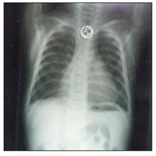
사진 205. 이물이 식도관 내에 있다.
Copyright ⓒ 2011 John Sangwon Lee, MD., FAAP
소화관 내 이물(인두 강 이물, 식도관 내 이물, 위 내 이물, 장관 내 이물/위장관 내 이물)의 치료
- 이물이 식도관 속이나 위 속으로 들어갔을 때 숨을 제대로 쉬고 의식이 있으면 의료구급대, 병원 응급실 또는 단골 의사에게 전화해서 그들의 지시에 따라 치료를 현장에서 시작한다.
- 이물이 식도관 속이나 위 속으로 들어갔을 때도 가능한 의사의 지시에 따라 응급 치료를 시작하는 것이 원칙이다.
- 의사의 지시를 곧 받을 수 없고 의식이 있고 숨을 정상적으로 쉴 수 있을 때는 얼굴을 바닥에 대고 옆으로 엎드려 누운 자세로 구급차로 병원 응급실로 급히 데리고 가는 것이 상책이다.
- 식도관 속에 걸려있고 더 이상 위 속 쪽으로 내려가지 않는 식도관 속 이물은 이비인후과 전문의, 흉곽외과 전문의 또는 그 외 다른 과 전문의가 식도내시경을 이용해 풍선 의료기구나 리트리벌 넷(Retrieval net) 의료기구 등으로 꺼낼 수 있다.
- 소화관 내에 있는 납 성분 이물은 가능한 속히 제거 해 내야한다.
- 식도관 속에 걸려있는 이물을 위 속 쪽으로 더 내려 보내기 위해 음식물을 강제로 먹여서는 안 된다.
- 식도 내 이물과 먹은 음식물이 식도관 속에 차 있을 수 있기 때문이다.
- 이런 경우 식도경 검사로 식도관 속 이물을 꺼낼 때 식도관 속을 차단한 이물과 먹었던 음식물을 동시 꺼내야 하기 때문에 치료 문제가 더 복잡해질 수 있다.
- 또 이때 먹은 음식물을 구토할 수 있고 구토물이 기도 속으로 흡인될 수 있다.
- 따라서 식도관 속에 이물이 있다고 의심되면 의사의 지시가 있기 전 절대로 음식물을 먹어서는 안 된다.
- 식도관 속 이물이 식도관에 인접된 기관이나 기관지의 일부를 눌러 호흡곤란이 생길 수 있다.
- 이런 때도 환아를 안정시키고 상체를 하체보다 15도 정도 낮게 옆으로 눕히고 의료구급대, 병원 응급실 또는 단골 의사의 지시에 따라 병원 응급실로 빨리 데리고 가야 한다.
- 동전이나 단추와 같이 표면이 둥글고 매끈매끈한 식도관 속 이물은 전체 식도관 속을 무난히 통과한 후 위 속으로 무난히 들어갈 수 있는 것이 보통이고 그 다음 그 이물이 소대장관 속을 무난히 지나서 대변과 같이 체외로 나오는 것이 보통이다.
- 이런 소화관 내 이물은 식도관 속으로 삼킨 후 2∼3일 이내에 대변으로 나오는 것이 보통이다.
- 그러나 핀, 열린 상태의 안전핀, 못, 바늘 등과 같이 끝이 뾰족한 이물은 식도관 속을 통과해서 위 속으로 들어간 다음 위 속에서 소장관 속으로 더 이상 내려가지 않고 거기에 그대로 머물러 있을 수 있다.
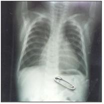
사진 206. 옷핀 이물이 위 속에 있다.
Copyright Ⓒ 2011 John Sangwon Lee, MD., FAAP
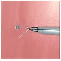
사진 207. 위 속 옷핀 이물을 내시경을 통해 육안으로 보고 내시경 튜브 속에다 넣고 꺼낼 수 있다.Copyright ⓒ 2011 John Sangwon Lee, MD., FAAP
- 이런 종류의 식도관 속 이물이나 위 속 이물은 자석이 달린 내시경을 이용해서 꺼낼 수 있다. 드물게는 위장관 속 이물을 수술해서 꺼내기도 한다.
- 대개, 겉이 매끈매끈하고 둥근 이물이 위장 속에 있을 때는 평소처럼 음식물을 먹어도 된다. 이 때 이물이 위 속을 통과한 후 소장관 속과 대장관 속을 지나가는 과정을 X-선 사진 검사로 알아볼 수 있다.
- 그렇지만 동전같이 표면이 둥글고 매끈거리는 이물이 위장관 속에 있을 때는 X-선 검사를 꼭 할 필요가 거의 없다.
- 끝이 날카로운 이물이 위장관 속에 있고 구토, 복통, 열 등의 증상 징후가 생기고 복막염 등이 생겼다고 의심되면 그에 따른 진단 치료를 적절히 한다.
- 카메라, 계산기, 시계 등에 들어 있는 디스크 배터리(디스크 전지)를 삼켜 식도관 속이나 위장관 속에 걸쳐 식도관 속 이물이 될 수 있고 위장관 속에 걸쳐 위장관 속 이물이 될 수 있다.
- 특히 식도관 속에 걸쳐 있는 디스크 전지는 불과 2-4시간 내에 식도관 점막층에 화상을 입힐 수 있고 심하면 식도관의 벽이 천공될 수 있다.
- 그런 이유로 디스크 전지가 위장관 속에 있을 때는 응급으로 제거 치료한다. 내시경 튜브 제거치료를 할 수 있다.
|
다음은“이물을 삼켰어요–반지를 삼켰어요 ”에 관한 인터넷 소아청소년 건강상담 질의응답의 예 입니다. |
Q&A. 이물을 삼켰어요–반지를 삼켰어요
Q.
7세 된 여아인데 지금 막 반지를 삼켰습니다.
어찌해야할지…..
억지로 토를 시켜보기도 했으나 소용이 없고 특별히 어디가 아프다거나 불편하다고는 하지 않는데 어찌해야 할지 걱정이 많이 됩니다.
가능하시면 빨리 답을 받아볼 수는 없는지요?
빠른 답변 부탁드려도 괜찮을까요?
A.
하늘님
다음은 www.koreapediatrics.com 홈페이지에 있는 소화관 이물에 관한 글입니다. 참고하시기 바랍니다.
[입안에 있던 구슬, 동전, 땅콩, 반지 등 이물은 입 밖으로 뱉어내든지 입안에 들어 있든지 인두, 인후, 기관, 기관지 등의 기도 내로 들어가든지 또는 인두, 식도, 위, 소대장관 등 위장관 내로 들어갈 수 있다.
입안에 있던 이물이 기도 내로 들어가면 들어갈 때 기침, 호흡곤란, 질식 등의 증상 징후가 생기는 것이 보통이고 때로는 기도가 완전히 막혀 질식해서 사망할 수 있고 기도의 일부분이 막힐 때는 호흡곤란이 생길 수 있습니다.
이물이 식도관 속으로 들어갈 때는 식도관 속으로 들어가기 바로 전 인두강과 식도관이 연결되는 식도관의 최상부에 있든지 그 이물이 식도관 속으로 좀 더 들어가서 식도관 속에 있든지 식도관 속을 통과한 후 위 속이나 소대장관 속에 들어가 있던지 또는 인두 강, 식도관, 위, 소대장관 속 등 위장관 속을 완전히 통과해서 대변으로 나올 수 있다.
이물이 인두 강 내에서 식도관 속으로 들어갈 때 이물질의 크기와 종류에 따라 기침을 할 수 있고 앞가슴이 뻐근하며 아플 수 있고 그 후 그 이물로 인하여 아무런 증상 징후 없이 위장관 속을 통과해서 변으로 나올 수 있다.]
이상 말씀드린 바와 같이 반지 등 식도관 속이물이나, 위장관 속 이물은 대변으로 자연적으로 나올 가능성이 많습니다.
또 그 반지가 기도 내로 들어가 있을 가능성도 있습니다.
아직까지 반지가 대변으로 나오지 않았으면 목, 가슴, 배 X-선 검사로 해보시기 바랍니다.
기도 내로 이물이 들어갈 때 기도가 일부 막힐 수 있습니다.
기도 내로 이물이 들어갔을 때 첫 1~2분 동안은 상당히 중요합니다.
그런 안전사고가 생기지 않게 평소에 예방하고 또 기도 내에 이물이 들어갔을 때 즉각 치료하는 방법을 평소에 배워야 합니다.
[부모도 반의사가 되어야 한다–소아가정간호백과]-제1권 소아청소년 응급의료–기도 속에 이물이 들어갔을 때.
제2권 소아청소년 질병 및 안전사고 예방–우발사고. 그리고 소화기 내에 이물이 들어갔을 때 등을 참조하시기 바랍니다.
소아청소년과에서 진찰 진단 치료를 받고 상담하시기 바랍니다. 질문이 더 있으시면 다시 연락 주세요. 감사합니다. 이상원 드림
Foreign bodies in digestive tracts 소화관 내 이물
Figure 202. Foreign body in the digestive tract a-a foreign body in the pharyngeal cavity, b, c, d-a foreign body in the esophagus, e- a foreign body in the stomach, f-a foreign body in the small intestine Source-Used with permission from Galaxo Wellcome
Figure 203. Foreign body in the digestive tract a, b, c- foreign body in the esophagus, d- foreign body in the stomach, e- foreign body in the small intestine Copyright ⓒ 2011 John Sangwon Lee, MD., FAAP
Foreign body in the digestive tract
• A foreign body in the pharyngeal cavity, a foreign body in the esophagus, a foreign body in the stomach, and a foreign body in the small intestine are collectively referred to as a foreign body in the digestive tract.
• According to statistics, 33% of foreign bodies in the digestive tract and foreign bodies in the airways of children and adolescents were foreign bodies in the trachea and foreign bodies in the bronchi, and 56% were foreign bodies in the esophagus and other foreign bodies in the digestive tract.
• Most of the foreign objects in the digestive tract such as foreign bodies in the pharyngeal cavity, foreign bodies in the esophagus, foreign bodies in the stomach (foreign bodies in the stomach), foreign bodies in the small intestine, foreign bodies in the colon, etc. were coins, buttons, small metal lumps, safety pins, etc.
• Most of the foreign objects in the airways were peanuts, beans, popcorn, pins, screws, wires, nails, and pencil erasers. It can become a foreign object in the esophageal tract by swallowing the disk battery (disk battery) contained in cameras, calculators, and watches, and it can become a foreign object in the gastrointestinal tract inside the stomach or in the small intestine. • Most of the foreign body in the airways occurs most frequently in infants under 5 years of age, and most of the foreign body in the digestive tract is said to occur most in infants and young children between 6 months and 3 years old.
• To prevent infants and toddlers from accidentally swallowing foreign objects and entering into the airways or digestive tract, take special care and prevent them in advance.
• The inside of the mouth, pharynx, esophagus, stomach, duodenum, small intestine, and large intestine belong to the digestive system (digestive system/digestive system), and the foreign body formed therein is called a foreign body in the digestive tract or a foreign body in the digestive tract.
Table 8. Foreign bodies in the digestive tract of children 표 8. 소아들의 소화관 내 이물
Types of foreign bodies in the digestive tract
| Types of foreign bodies in the digestive tract | Foreign bodies in the digestive tract |
| Foods | Fruit seeds such as peach seeds, pieces of meat bones, fish spines or bones, etc. |
| wood | Accessories such as wooden pieces of wood, toothpicks, wooden toys, etc |
| Batteries | Camera batteries, disk batteries such as calculators and watches (disc batteries), etc. |
| Jewelry | Glass pieces, earrings, rings, etc. |
| Cosmetic | pen cap, pencil, rubber eraser, crayon, paper clip, etc |
| Toy | beads, air pen caps, etc. |
| Other | coins, needles, pins, hairballs in the stomach, etc. |
Symptoms, signs of a foreign body in the digestive tract (a foreign body in the pharyngeal cavity, a foreign body in the esophageal tract, a foreign body in the stomach, a foreign body in the intestine/a foreign body in the gastrointestinal tract)
Figure 204. Foreign objects in the esophagus or gastrointestinal tract can be trapped in the physiologically narrow esophagus and in the gastrointestinal tract. Figures a, b, c, e, f, g. a-cricoid pharynx, b-aortic arch, c-cardinal, e-pyloric, f-tris ligament, g-chief of the cemetery. CopyrightⒸ 2011 John Sangwon Lee, MD., FAAP
• Infants and toddlers are curious and inexperienced in making judgments, so they have a habit of sucking and playing with almost anything small enough to fit in their mouth. • Putting it in your mouth, drinking, sucking, and playing can accidentally get into your airways or digestive tract.
• Most of the foreign matter that has entered the pharyngeal cavity passes through the pharynx, passes through the esophagus and gastrointestinal tract, and comes out of the anus without problems.
• Otherwise, it passes through the pharyngeal cavity, the esophagus, and the gastrointestinal tract, and depending on the type of the foreign object, it does not go down over one part of the pharyngeal cavity or the gastrointestinal tract and becomes a foreign object in the digestive tract.
• Symptoms and signs of a foreign body in the digestive tract differ depending on various conditions, such as where the foreign body is in the digestive tract. • Depending on the foreign body, when the foreign body enters the esophageal tract from the pharyngeal cavity, it may be whacking, unable to breathe for a while, drooling, and stiff chest.
• When a foreign object with a round surface, such as a coin or a gonggi, passes through the pharyngeal cavity and enters the esophagus, it may cause a slight coughing after a while.
• Sometimes, you may feel that the foreign body enters the upper part of the esophageal tract and then gradually and slowly descends toward the stomach.
• Most of the foreign bodies in the esophagus go into the stomach and then go through the small and large intestine within 2 to 5 days and then come out of the anus like feces.
• Some parents brought their infants and children to the Department of Pediatrics and adolescents and said,’The child was playing with something in his mouth, then suddenly picked up a case, coughed, had a sore throat, breathed for a while, and now it seems okay. I don’t know if it got into my throat.’
• When a foreign body enters the esophageal tract, breathing difficulties may occur at first, almost similar to when a foreign body enters the trachea or bronchi, but shortly after the foreign body enters the esophagus through the pharyngeal cavity, the dyspnea is much milder. It is usually not happening.
• Before the foreign body passes through the esophagus and enters the stomach, the anterior chest may stiffen. There are times when a foreign body in the esophageal canal gets caught in a part of the esophageal canal and does not go down any further.
• At this time, a part of the esophageal canal may be completely or incompletely blocked.
• Symptoms may vary slightly depending on the degree of blockage.
• If the inside of the esophagus is completely blocked, you can drool a lot, and the food you eat can pass through the esophagus under the blocked esophagus and go down to the stomach, and your anterior chest may be sore.
• It is common to vomit because the food you have eaten can no longer go down into your stomach in the esophagus.
• Rarely, after a very small foreign body enters the esophageal canal, it may remain there for a long time, leaving a portion of the esophageal canal incompletely blocked, and no symptoms may appear.
• Most of the small foreign objects with sharp tips such as open safety pins, nails, or needles pass well into the esophagus, then pass safely through the stomach and the small intestine, and come out of the antigen like feces within 2 to 3 days.
• Certain foreign objects with pointed tips may become lodged in the esophageal wall or the gastrointestinal wall mucosa when passing through the esophagus or gastrointestinal tract, causing damage to the esophageal or gastrointestinal wall.
• When this type of foreign body enters the digestive tract, it can be diagnosed with a chest x-ray or abdominal x-ray to determine where the foreign body is in the digestive tract.
• This type of foreign body can damage the esophagus or gastrointestinal tract and cause peritonitis or other complications.
• Perforation in the wall of the gastrointestinal tract caused by a foreign body can cause peritonitis, which can lead to abdominal pain, vomiting, and fever.
• A small disk cell can perforate the esophageal canal wall after about 2-4 hours or more over the esophageal canal. Since there is a possibility that foreign bodies in the esophageal tract of infants older than 6 years of age with a diameter of about 15 mm may not naturally descend into the gastrointestinal tract, treatment to remove the foreign bodies early is required.
Diagnosis of a foreign body in the digestive tract (a foreign body in the pharynx, a foreign body in the esophageal tract, a foreign body in the stomach, a foreign body in the intestine / a foreign body in the gastrointestinal tract)
• The alimentary canal or tract is the organ from the mouth to the esophagus, stomach, duodenum, small intestine, large intestine, and anus.
• If it is suspected that a foreign body has entered the digestive tract by synthesizing symptoms and examination findings, an X-ray picture of the mouth, neck, chest, and stomach can be taken to diagnose a foreign body in the digestive tract (see photo 205).
• X-rays can almost determine what part of the digestive tract is in the digestive tract and what kind of foreign matter is, but X-rays when a foreign object from a toy or object made of some kind of plastic is in the digestive tract or airways. It may not appear well on photo inspection.
• Sometimes this type of foreign body in the digestive tract cannot be reliably diagnosed with an x-ray.
• Sometimes it is not possible to reliably determine that there is no foreign body in the digestive tract or in the respiratory tract, even though the x-rays that were examined to diagnose a foreign body in the digestive tract or airway did not show the foreign body.
• If there are persistent signs of any symptoms that may be caused by a foreign body in the digestive tract or a foreign body in the airways, the diagnosis may be performed by tests other than an X-ray examination.
Picture 205. The foreign body is in the esophagus. Copyright ⓒ 2011 John Sangwon Lee, MD., FAAP
Treatment of a foreign body in the digestive tract (a foreign body in the pharyngeal cavity, a foreign body in the esophageal tract, a foreign body in the stomach, a foreign body in the intestine / a foreign body in the gastrointestinal tract)
• When a foreign object enters the esophagus or stomach, breathe properly and, if conscious, call a medical paramedic, hospital emergency room, or a regular doctor and begin treatment on-site according to their instructions.
• When a foreign body enters the esophagus or stomach, it is a rule to start emergency treatment according to the doctor’s instructions as much as possible.
• If you cannot get directions from your doctor soon, and you are conscious and can breathe normally, it is best to take it to the hospital emergency room by ambulance in a lying position with your face on your side.
• For foreign objects in the esophagus that are trapped in the esophagus and do not go down to the stomach anymore, an otolaryngologist, thoracic surgeon, or another surgeon can use a balloon medical device or Retrieval net medical treatment using an esophageal endoscopy. Can be taken out with utensils, etc.
• Lead-based foreign matter in the digestive tract should be removed as soon as possible.
• Food should not be forcibly fed in order to further lower the stomach into foreign objects trapped in the esophageal tract.
• This is because foreign objects and food in the esophagus may be filled in the esophagus.
• In this case, the treatment problem may be more complicated because the foreign body blocked in the esophagus and the food eaten must be removed at the same time when the foreign body is removed from the esophagus through an esophageal examination.
• You may also vomit the food you eat at this time and the vomit may be sucked into your airways.
• Therefore, if you suspect you have a foreign body in your esophagus, you should never eat food before your doctor tells you to.
• A foreign body in the esophageal canal may press on a part of the trachea or trachea adjacent to the esophageal canal, causing dyspnea.
• Even in such a case, you should stabilize the child, lay the upper body 15 degrees lower than the lower body, and take him to the hospital emergency room as directed by a medical paramedic, hospital emergency room, or regular doctor.
• Foreign objects in the esophagus, which have a round and smooth surface, such as coins or buttons, can pass through the entire esophagus and then enter the stomach. Then, the foreign object passes through the platoon and is outside the body like stool. It is common to come out as.
• These foreign bodies in the digestive tract usually come out of the stool within 2 to 3 days after being swallowed into the esophagus.
• However, foreign objects with pointed tips such as pins, safety pins in an open state, nails, needles, etc. may pass through the esophagus and enter the stomach and then remain there without further descending into the small intestine from the stomach.
Picture 206. The clothespin foreign body is in the stomach. Copyright Ⓒ 2011 John Sangwon Lee, MD., FAAP
Photo 207. Clothespins inside the stomach can be seen with the naked eye through an endoscope, and inserted into the tube of the endoscope. Copyright © 2011 John Sangwon Lee, MD., FAAP
• This type of foreign body in the esophagus or stomach can be removed using an endoscope with a magnet. Rarely, a foreign body in the gastrointestinal tract is surgically removed.
• In general, you can eat food as usual when the outside is smooth and there is a round foreign body in your stomach. At this time, the process of passing through the inside of the small intestine and the inside of the colon can be seen by X-ray examination.
• However, when there is a foreign object in the gastrointestinal tract with a round and smooth surface like a coin, it is rarely necessary to perform an X-ray examination.
• If a foreign object with a sharp tip is in the gastrointestinal tract, symptoms such as vomiting, abdominal pain, fever, and peritonitis are suspected, appropriate diagnostic treatment is required.
• Swallowing disk batteries (disc batteries) contained in cameras, calculators, watches, etc., may become foreign objects in the esophagus or gastrointestinal tract, or foreign objects in the gastrointestinal tract throughout the gastrointestinal tract.
• Disc cells, especially in the esophageal canal, can burn the mucous membrane of the esophageal canal in just 2-4 hours, and in severe cases, the walls of the esophageal canal can be perforated.
• For that reason, if the disk battery is in the gastrointestinal tract, it should be removed and treated as an emergency. Endoscopic tube removal treatment can be performed.
The following is an example of an Internet pediatric and adolescent health counseling question and answer for “I swallowed a foreign body-I swallowed a ring”.
Q&A. I swallowed a foreign body-I swallowed a ring
Q.
I’m a 7-year-old girl, and I just swallowed the ring. What to do… She even tried to force her to vomit, but to no avail, and she doesn’t particularly say that she’s sick or uncomfortable, but she’s worried a lot about what to do. Can’t you get an answer as soon as possible? Would it be okay to ask for a quick answer? A. Heaven The following is an article on foreign bodies in the digestive tract on the website of www.koreapediatrics.com.
Please note. [Foreign substances such as beads, coins, peanuts, rings, etc. in the mouth are spit out of the mouth or enter the airways such as the pharynx, throat, trachea, and bronchi, or enter the gastrointestinal tract such as the pharynx, esophagus, stomach, and small intestine.
I can. When a foreign body in the mouth enters the airways, symptoms such as coughing, shortness of breath, and suffocation are common when entering. Sometimes, the airways are completely blocked, resulting in suffocation and death, and when a part of the airways is blocked, breathing difficulties may occur.
When a foreign body enters the esophageal canal, whether it is at the top of the esophageal canal that connects the pharyngeal cavity and the esophageal canal immediately before entering the esophageal canal, the foreign body enters the esophageal canal and passes through the esophageal canal. The posterior stomach or the platoon may pass through the gastrointestinal tract, such as the pharyngeal cavity, the esophagus, the stomach, or the platoon, and come out as feces.
When a foreign body enters the esophageal tract from the pharyngeal cavity, depending on the size and type of the foreign body, it may cause coughing, and the front chest may be stiff and painful, and then the foreign body may pass through the gastrointestinal tract without any symptomatic symptoms and come out into the stool. have.] As mentioned above, foreign objects in the esophageal tract, such as rings, or foreign objects in the gastrointestinal tract, are likely to come out naturally as feces. It is also possible that the ring is in the airway. If the ring has not yet come out in your stool, try an x-ray of your neck, chest, and stomach. When a foreign object enters the airway, the airway may be partially blocked.
It is very important for the first 1-2 minutes when a foreign object enters the airway. To prevent such safety accidents, you should learn how to prevent them and treat them immediately when a foreign body gets into the airways.www.drleepediatrics.com-Vol. 1 Emergency medical care for children and adolescents-When a foreign body enters the airway. Volume 2 Prevention of Child and Adolescent Diseases and Safety Accidents-Accidents. Also, please refer to when a foreign object gets into the fire extinguisher. Please consult with the Department of Pediatrics and Adolescents after receiving medical examination and treatment. If you have more questions, please contact us again. Thank you. Lee Sang-won.
• Even at such a time, the patient is stabilized and the upper body is more than the lower body.
출처 및 참조 문헌 Sources and references
- NelsonTextbook of Pediatrics 22ND Ed
- The Harriet Lane Handbook 22ND Ed
- Growth and development of the children
- Red Book 32nd Ed 2021-2024
- Neonatal Resuscitation, American Academy Pediatrics
- www.drleepediatrics.com제7권 소아청소년 감염병
- www.drleepediatrics.com제9권 소아청소년 소화기 질환
- Red book 29th-31st edition 2021
- Nelson Text Book of Pediatrics 19th — 21st Edition
- The Johns Hopkins Hospital, The Harriet Lane Handbook, 22nd edition
-
Childhood Emergencies in the Office, Hospital and Community, American Academy of Pediatrics
-
Emergency Medical Service for Children, By Ross Lab. May 1989. p.10
-
Emergency care, harvey grant and robert murray
-
Emergency Care Transportation of Sick and Injured American Academy of Orthopaedic Surgeons
-
Emergency Pediatrics A Guide to Ambulatory Care, Roger M. Barkin, Peter Rosen
-
Immediate care of the acutely ill and injured, Hugh E. Stephenson, Jr
-
The Critically Ill Child, Diagnosis and Management, Edited by Clement A. Smith
-
Emergency Medical Services for Children: The Role of the Primary Care Provider, America Academy of Pediatrics
-
Quick Reference To Pediatric Emergencies , Delmer J. Pascoe, M.D., Moses Grossman, M.D. with 26 contributors
-
Manual of Emergency Care
-
응급환자관리 정담미디어
-
소아가정간호백과–부모도 반의사가 되어야 한다, 이상원
-
Neonatal Resuscitation American heart Association
-
Neonatology Jeffrey J.Pomerance, C. Joan Richardson
-
Pediatric Resuscitation Pediatric Clinics of North America, Stephen M. Schexnayder, M.D.
-
Pediatric Critical Care, Pediatric Clinics of North America, James P. Orlowski, M.D.
-
Preparation for Birth. Beverly Savage and Dianna Smith
- Infectious disease of children, Saul Krugman, Samuel L Katz, Ann A. Gershon, Catherine Wilfert
-
The Harriet Lane Handbook 19th Edition
-
소아과학 대한교과서
-
제1권 소아청소년 응급의료 참조문헌과 출처
-
Other
Copyright ⓒ 2015 John Sangwon Lee, MD., FAAP
“부모도 반의사가 되어야 한다”-내용은 여러분들의 의사로부터 얻은 정보와 진료를 대신할 수 없습니다.
“The information contained in this publication should not be used as a substitute for the medical care and advice of your doctor. There may be variations in treatment that your doctor may recommend based on individual facts and circumstances. “Parental education is the best medicine.”