상완골 골절, 척골 골절, 요골 골절, 팔꿈치 관절 골절 Fracture of humerus, radius, ulna, elbow joint
상완골 골절, 척골 골절, 요골 골절, 팔꿈치 관절 골절의 원인
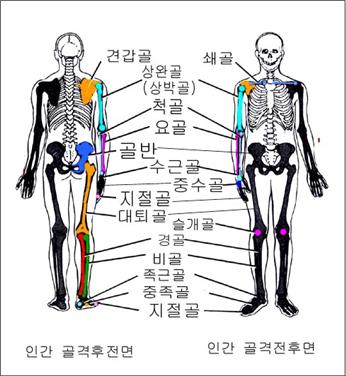
그림 112. 인간 골격 후면, 전면
출처-Emergency Care, By Grant And Murray
-
어깨 관절(견갑 관절/견관절)과 팔꿈치 관절(주관절)을 연결하는 뼈를 상완골 또는 상박골이라고 한다.
-
운동, 교통사고, 또는 안전사고 등으로 상박골이 골절될 수 있다.
-
손과 아래팔을 쭉 뻗은 상태에서 잘못 넘어져 상완골 골절이 생기면 상완골의 위 부위가 골절되기 쉽다.
-
상완골 골두에 골절이 생길 수 있고 골간 골절이 생길 수 있고 골단 골절이 생길 수 있다.
-
그 중 어느 부위의 상완골이 골절됐느냐에 따라 증상 징후가 다르고 치료도 다르다.
-
상완골의 중간 부위가 강타 당했을 때는 상완골의 중간 부위가 골절되기 쉽고,
-
팔꿈치를 짚고 넘어질 때는 팔꿈치 부위 상완골이 골절되기 쉽다.
-
상완골이 골절될 때 골절된 상완골 부위 주위에 있는 혈관 및, 또는 말초신경 등이 손상될 수 있다.
-
이와 같이 상완골이 여러 형태로 골절될 수 있다.
-
상완골 골절 이외 다른 골절에 관한 정보는 “골절(Fractures)” 참조
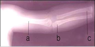
사진 113. 유아 상완골(a), 척골(b), 요골(c)의 x선 사진
Copyright ⓒ 2013 John Sangwon Lee, M.D., FAAP
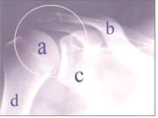
사진 114. 오른 쪽 견관절(어깨 관절)의 X-선 사진
○–어깨 관절, a-상완골 두부, b-쇄골, c-견갑골, d-상완골 골간
Copyright ⓒ 2013 John Sangwon Lee, M.D., FAAP
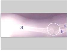
사진 115. 유아의 상완골, 척골, 요골의 X-선 사진
○–팔꿈치 관절(주관절), a-상완골, b-척골, c-요골
Copyright ⓒ 2013 John Sangwon Lee, M.D., FAAP
상완골 골절, 척골 골절, 요골 골절, 팔꿈치 관절 골절의 증상 징후
-
상완골의 어느 부위에 골절됐는지, 골절된 정도에 따라 증상 징후가 다르다.
-
골절과 함께 생긴 외상의 종류와 정도에 따라 부수되는 증상 징후가 다르다.
-
골절된 위팔 부위가 붓고 아프고 그 쪽 팔이 능동적으로나 수동적으로 움직일 때는 상완골 골절이 있는 위 팔 부위가 더 아프고 때로는 움직일 수 없다.
-
골절된 위 팔 부위를 손으로 누르면 아프다.
-
그 위 팔을 능동적으로 움직이기를 싫어하고 정상적으로 움직일 수 없는 때가 많다.
-
상완골 골절의 끝으로 그 주위에 있는 말초신경이나 혈관 또는 근육 등이 손상될 수 있다. 그 쪽 팔에 있는 말초신경이 손상되면 그 말초신경이 컨트롤하는 근육들의 일부가 마비될 수 있다.
-
이때 마비된 신경의 종류와 그의 분포에 따라 그 쪽 위팔을 능동적으로 움직일 수도 없고 그 쪽 아래팔의 일부나 전체의 감각에 이상이 생길 수 있다.
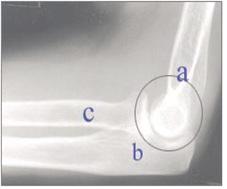
사진 116. 소아의 정상 상완골, 척골, 요골, 팔꿈치 관절의 X-선 사진
◯내–팔꿈치 관절, a-상완골, b-척골, c-요골
Copyright ⓒ 2013 John Sangwon Lee, M.D., FAAP

사진 117. 상완골 골절 X-선 사진
◯내–상완골 골절, a-상완골, b-척골, c-요골
Copyright ⓒ 2013 John Sangwon Lee, M.D., FAAP

사진 118. 정상 팔꿈치 관절(○ 내)
Copyright ⓒ 2011 John Sangwon Lee, M.D., FAAP
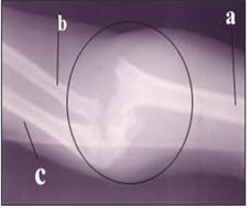
사진 의료. 심한 팔꿈치 관절 골절(◯ 내)
Copyright ⓒ 2011 John Sangwon Lee, M.D., FAAP
- 그뿐만 아니라 상관골 골절이 생긴 부위에 있는 혈관이 손상되면 그쪽의 위 아래 팔의 혈액순환이 잘 되지 않아 그쪽의 위아래 팔의 피부색이 파래지거나 손톱 밑 색도 파라질 수 있다.
상완골 골절, 척골 골절, 요골 골절, 팔꿈치 관절 골절의 진단
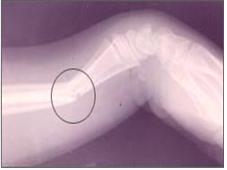
사진 121. ◯ 내 척골과 요골에 생긴 복합 골절
Copyright ⓒ 2013 John Sangwon Lee, M.D., FAAP
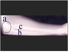
사진 120. 정상 팔꿈치 관절, 척골과 요골의 X-선 사진
◯–팔꿈치 관절, a-상완골, b-척골, c-요골
Copyright ⓒ 2013 John Sangwon Lee, M.D., FAAP
-
병력 증상 징후와 진찰소견 등을 종합해서 상완골 골절이 생겼고 의심되면
-
상완골 X-선 사진을 찍어 확진할 수 있다.
상완골 골절, 척골 골절, 요골 골절, 팔꿈치 관절 골절의 치료
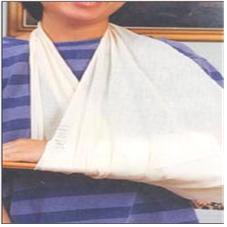
사진 122. 경미한 척골 골절이나 요골 골절이 의심되면 의사의 지시에 따라 그쪽 앞 팔에 부목을 대고 삼각건으로 받친다.
Copyright ⓒ 2011 John Sangwon Lee, M.D., FAAP
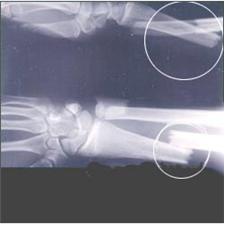
사진 123. 심한 척골 골절과 요골 골절 X-선 사진(상◯내, 하◯내)
Copyright ⓒ 2011 John Sangwon Lee, M.D., FAAP
-
가능한 한 환아를 안정시키고 안전한 곳으로 옮긴다.
-
상완골 골절의 정도에 따라 의료구급대원이나 병원 응급실이나 의사에게 응급 전회 진료 상담을 해 그들의 지시에 따라 현장에서 치료를 시작한다.
-
상완골 골절이 있는 쪽의 팔과 손의 혈액순환이 정상인지, 말초신경 마비가 되어 있는지를 가능하면 빨리 체크해 본다.
-
상황에 따라 의사나 구급대원이 현장에 도착하기 전에는 가능한 한 골절된 위팔을 처음 본 상태 그대로 유지하고 상완골 골절이 된 위아래 팔을 더 이상 움직이지 말아야 한다.
-
경미한 상완골 골절이 있는 쪽 위팔, 아래팔, 손목, 손을 함께 삼각건의 한쪽으로 받쳐주면서 삼각건의 다른 쪽을 목에다 걸쳐 응급으로 처치할 수 있다.
-
이 응급치료도 의사의 지시에 따라 한다.
-
골절된 위팔을 환아의 몸통에 대고 몸통과 골절된 위팔을 다른 삼각건으로 고정시키고 병원으로 갈 수 있다.
-
상완골 골절이 있는 위팔을 삼각건으로 받쳐 고정시킬 때 골절된 상완골을 밀거나 끌어 잡아당기거나 비틀어서는 안 된다.
-
가능한 한 처음 목격한 골절 상태 자세 그대로 계속 유지하면서 조심스럽게 삼각건으로 받쳐줄 수 있다.
-
병원에서는 골절된 상완골의 부위에 따라 골절의 정도에 따라 석고붕대 고정 치료나 견인치료, 또는 수술 치료로 치료한다.
Fracture of humerus, radius, ulna, elbow joint Causes of humerus fractures, ulnar fractures, radial fractures, elbow joint fractures 상완골 골절, 척골 골절, 요골 골절, 팔꿈치 관절 골절
Figure 112. Human skeleton back and front Source – Emergency Care, By Grant And Murray
• The bone that connects the shoulder joint (shoulder joint/shoulder joint) and the elbow joint (elbow joint) is called the humerus or humerus.
• The humerus can be fractured due to sports, traffic accidents, or safety accidents.
• If a humerus fracture occurs due to an accidental fall while the hand and forearm are outstretched, the upper part of the humerus is easily fractured.
• Fractures of the humeral head may occur, interosseous fractures may occur, and epiphyseal fractures may occur.
• Symptoms and treatment differ depending on which part of the humerus is fractured.
• When the middle part of the humerus is struck, it is easy to fracture the middle part of the humerus,
• When you fall on your elbow, the humerus in the elbow area is easy to fracture. • When the humerus is fractured, blood vessels and/or peripheral nerves around the fractured humerus may be damaged.
• The humerus can be fractured in many ways.
• See “Fractures” for information on fractures other than humerus fractures.
Picture 113. X-ray of infant humerus (a), ulna (b), and radius (c) Copyright ⓒ 2013 John Sangwon Lee, M.D., FAAP
Picture 114. X-ray of the right shoulder joint (shoulder joint) ○-shoulder joint, a-humerus head, b-clavicle, c-scapula, d-humerus shaft Copyright ⓒ 2013 John Sangwon Lee, M.D., FAAP
Picture 115. X-rays of the humerus, ulna, and radius of an infant ○-elbow joint (elbow joint), a-humerus, b-ulnar, c-radius Copyright ⓒ 2013 John Sangwon Lee, M.D., FAAP
Symptom, signs of humerus fracture, ulnar fracture, radial fracture, elbow joint fracture
• Symptoms vary depending on where the fracture of the humerus is and the extent of the fracture.
• Signs of concomitant symptoms differ depending on the type and severity of the trauma associated with the fracture.
• When the fractured upper arm is swollen and painful and the arm is actively or passively moved, the upper arm with the humerus fracture is more painful and sometimes unable to move. • It hurts when you press your hand on the fractured upper arm.
• He does not like to actively move his upper arm and is often unable to move normally.
• At the end of the humerus fracture, peripheral nerves, blood vessels, or muscles around it may be damaged. Damage to a peripheral nerve in that arm can paralyze some of the muscles it controls.
• At this time, depending on the type and distribution of the paralyzed nerve, the upper arm may not be able to move actively, and a part or all of the sense of the forearm may be abnormal.
Picture 116. X-rays of normal humerus, ulna, radius, and elbow joints in a child ◯Inner-elbow joint, a-humerus, b-ulnar, c-radius Copyright ⓒ 2013 John Sangwon Lee, M.D., FAAP
Picture 117. X-ray of humerus fracture ◯Intra-humerus fracture, a-humerus, b-ulnar, c-radius Copyright ⓒ 2013 John Sangwon Lee, M.D., FAAP
Picture 118. Normal elbow joint (within ○) Copyright ⓒ 2011 John Sangwon Lee, M.D., FAAP
Photo medical. Severe elbow joint fracture (within ◯) Copyright ⓒ 2011 John Sangwon Lee, M.D., FAAP
• In addition, if the blood vessels in the area of the fracture of the superior cortex are damaged, the blood circulation in the upper and lower arms is not good, and the skin color of the upper and lower arms may turn blue or the color under the nails may also become pale.
Diagnosis of humerus fractures, ulnar fractures, radial fractures, and elbow joint fractures
Picture 121. ◯ Compound fractures in my ulna and radius Copyright ⓒ 2013 John Sangwon Lee, M.D., FAAP
Picture 120. X-ray picture of normal elbow joint, ulna and radius ◯ – elbow joint, a – humerus, b – ulna, c – radius Copyright ⓒ 2013 John Sangwon Lee, M.D., FAAP
• If a humerus fracture has occurred and is suspected based on the medical history, symptoms, signs, and examination findings,
• Diagnosis can be confirmed by taking an X-ray of the humerus.
Treatment of humerus fractures, ulnar fractures, radial fractures, and elbow joint fractures
Photo 122. If a minor fracture of the ulna or radius is suspected, as directed by the doctor, place a splint on the front arm and support it with a triangular tendon. Copyright ⓒ 2011 John Sangwon Lee, M.D., FAAP
Picture 123. X-rays of severe ulnar fracture and radial fracture (inner upper, lower inner) Copyright ⓒ 2011 John Sangwon Lee, M.D., FAAP
• As much as possible, stabilize the child and move him to a safe place.
• Depending on the severity of the fracture of the humerus, consult a medical paramedic, hospital emergency room, or doctor for emergency round-the-clock consultation and start treatment on the spot according to their instructions.
• Check as soon as possible whether blood circulation in the arm and hand on the side of the humerus fracture is normal and whether there is peripheral nerve palsy.
• Depending on the circumstances, the fractured upper arm should remain as it was at first sight as far as possible and the upper and lower arms with a fractured humerus should not be moved any further before the doctor or paramedics arrive at the scene.
• First aid can be given by placing the upper arm, forearm, wrist, and hand on the side with a minor humerus fracture together on one side of the tendon while supporting the other side of the tendon over the neck.
• This first aid treatment should also be done as directed by your doctor. • You can place the fractured upper arm on the patient’s torso, fix the torso and the fractured upper arm with another triangle tendon, and go to the hospital.
• Do not push, pull or twist the fractured humerus when supporting and immobilizing the upper arm with a fractured humerus.
• As far as possible, you can carefully support the fracture with a triangular tendon while remaining in the position of the first fracture.
• In hospitals, depending on the fractured humerus, depending on the severity of the fracture, it is treated with fixed casts, traction therapy, or surgical treatment.
출처 및 참조 문헌 Sources and references
- NelsonTextbook of Pediatrics 22ND Ed
- The Harriet Lane Handbook 22ND Ed
- Growth and development of the children
- Red Book 32nd Ed 2021-2024
- Neonatal Resuscitation, American Academy Pediatrics
- www.drleepediatrics.com 제16권 소아청소년 정형외과 질환
- www.drleepediatrics.com제8권 소아청소년 호흡기 질환
- www.drleepediatrics.com제9권 소아청소년 소화기 질환
- www.drleepediatrics.com제10권. 소아청소년 신장 비뇨 생식기 질환
- www.drleepediatrics.com제11권. 소아청소년 심장 혈관계 질환
- www.drleepediatrics.com제12권. 소아청소년 신경 정신 질환, 행동 수면 문제
- www.drleepediatrics.com제13권. 소아청소년 혈액, 림프, 종양 질환
- www.drleepediatrics.com제14권. 소아청소년 내분비, 유전, 염색체, 대사, 희귀병
- www.drleepediatrics.com제15권. 소아청소년 알레르기, 자가 면역질환
- Red book 29th-31st edition 2021
- Nelson Text Book of Pediatrics 19th — 21st Edition
- The Johns Hopkins Hospital, The Harriet Lane Handbook, 22nd edition
-
Quick Reference to Pediatric Emergencies, Delmer J. Pascoe, M.D., p.160
-
Emergency Pediatrics, A guide to ambulatory care, 5th edi. Roger M. Barkin, Peter Rosen, p.524-532
-
Emergency care and transportation of the sick and injured, 3rd edition, American Academy of orthopedic surgeons. p.25, 148-155
-
Nelson textbook, 14 edition, p.457, 1723
-
Childhood Emergencies in the Office, Hospital and Community, American Academy of Pediatrics
-
Emergency Medical Service for Children, By Ross Lab. May 1989. p.10
-
Emergency care, Harvey grant, and Robert Murray
-
Emergency Care Transportation of Sick and Injured American Academy of Orthopaedic Surgeons
-
Emergency Pediatrics A Guide to Ambulatory Care, Roger M. Barkin, Peter Rosen
-
Immediate care of the acutely ill and injured, Hugh E. Stephenson, Jr
-
The Critically Ill Child, Diagnosis and Management, Edited by Clement A. Smith
-
Emergency Medical Services for Children: The Role of the Primary Care Provider, America Academy of Pediatrics
-
Quick Reference To Pediatric Emergencies, Delmer J. Pascoe, M.D., Moses Grossman, M.D. with 26 contributors
-
Manual of Emergency Care
-
응급환자관리 정담미디어
-
소아가정간호백과–부모도 반의사가 되어야 한다, 이상원
-
Neonatal Resuscitation American heart Association
-
Neonatology Jeffrey J.Pomerance, C. Joan Richardson
-
Pediatric Resuscitation Pediatric Clinics of North America, Stephen M. Schexnayder, M.D.
-
Pediatric Critical Care, Pediatric Clinics of North America, James P. Orlowski, M.D.
-
Preparation for Birth. Beverly Savage and Dianna Smith
- Infectious disease of children, Saul Krugman, Samuel L Katz, Ann A. Gershon, Catherine Wilfert
-
The Harriet Lane Handbook 19th Edition
-
소아과학 대한교과서
-
제1권 소아청소년 응급의료 참조문헌과 출처
-
Other
Copyright ⓒ 2015 John Sangwon Lee, MD., FAAP
“부모도 반의사가 되어야 한다”-내용은 여러분들의 의사로부터 얻은 정보와 진료를 대신할 수 없습니다.
“The information contained in this publication should not be used as a substitute for the medical care and advice of your doctor. There may be variations in treatment that your doctor may recommend based on individual facts and circumstances. “Parental education is the best medicine.”