동맥관 개존 Patent ductus arteriosus
동맥관 개존의 개요
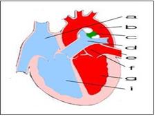
그림 30. 동맥관 개존(c)
a-우심방, b-대동맥, c-동맥관 개존, d-폐동맥, e-폐정맥, f-좌심방, g-좌심실, i-우심실
Copyright ⓒ 2013 John Sangwon Lee, MD., FAAP
-
출생 전 태아의 대동맥과 폐동맥 사이에 연결된 짤막한 혈관이 하나가 정상적으로 있다.
-
이 혈관을 동맥관이라고 한다(그림 10 태아의 혈액 순환도, 그림 30, 31, 32 참조).
-
출생 전 태아들의 정상 혈액순환은 출생 후 신생아들을 비롯한 영유아들, 학령기 아이들, 사춘기 아이들이나 성인들의 정상 혈액순환과 다른 점이 많다.
-
출생 전 태아들의 폐동맥과 대동맥을 연결하는 동맥관이 개존되어 있고 또 우심방과 좌심방 사이 심방 중격에 난원공이 정상적으로 있다.
-
태어난 후, 정상적으로 우심실 속 정맥혈의 전부가 폐동맥을 거쳐 폐 속으로 흘러간다.
-
그러나 태어나기 전 태아의 경우, 우심실 속에 있는 모든 혈액이 일단 폐동맥 속으로 흘러 가다가 그 혈액의 일부는 나머지 폐동맥 속을 계속 통과한 다음 마지막으로 폐 속으로 흘러가고
-
나머지 부분의 혈액은 동맥관을 통과해서 대동맥 속으로 흘러 들어가는 것이 정상 태아혈액 순환과정이다.
-
그렇지만 출생 후 바로 동맥관은 정상적으로 저절로 막히고 우심실 속 정맥혈의 전부가 폐동맥을 거처 폐 속으로 흘러간다. 이 때는 우심실에서 흘러온 폐동맥 속 혈액이 정상적으로 막힌 동맥관을 통과해서 대동맥 속으로 더 이상 흘러갈 수가 없다 (그림 10 태아 혈액 순환도 참조).
-
그러나 드물게, 출생 후 동맥관이 완전히 막히지 않고 계속 열려 있는 경우가 있다.
-
이런 혈관 기형을 동맥관 개존이라고 하고 그로 인해 생긴 증상 징후를 동맥관 개존증이라 한다.
그림 32. 동맥관 개존
◯로 표시된 부위에 있는 동맥관 개존
Copyright ⓒ 2013 John Sangwon Lee, MD., FAAP
그림 31. 동맥관 개존
◯로 표시된 부위에 있는 동맥관 개존
Copyright ⓒ 2013 John Sangwon Lee, MD., FAAP
동맥관 개존의 원인
-
원인은 아직 확실히 모른다.
-
임신 첫 3개월 기간에 임신부가 풍진을 앓을 때 태아도 풍진바이러스에 감염되면 풍진에 걸릴 수 있고 동맥관이 출생 후 계속 개존 될 수 있다.
-
또 특히 체중이 1,500∼1,750g 정도 되는 아주 작은 미숙 신생아들의 15%에서 출생 후 얼마 동안 동맥관이 일시적으로 개존되어 있을 수 있다.
-
출생 후 동맥관이 계속 개존되어 있을 때는 대동맥 속 동맥혈이 동맥관을 통과해서 폐동맥 속으로 흘러 들어간다.
-
태어나기 전과 정반대의 동맥관 혈액 순환 과정이 생기는 셈이다.
-
대동맥 속 혈액이 동맥관 속을 통과해서 흘러간 폐동맥 속 동맥혈과 우심실 속에서 폐동맥 속으로 흘러 들어온 정맥혈이 섞인 동맥혈, 정맥혈이 폐동맥 속을 통과 해 폐 속으로 흘러 들어간다.
-
따라서 동맥관 개존이 있을 때 폐가 정상 이상으로 더 많은 혈액 양을 취급해야 하는 부담을 갖게 된다.
-
이런 이유로, 심장은 정상 이상으로 더 많은 혈액 양을 펌프하게 되고 심장 전체가 보통 이상으로 더 많은 펌프 부담을 갖게 된다.
-
결국 좌심방과 좌심실이 비정상으로 더 커질 수 있고 좌 폐동맥 분지맥과 우 폐동맥 분지맥의 혈압이 비정상적으로 더 상승되고 그에 따른 여러 가지의 증상 징후가 생길 수 있다.
동맥관 개존의 증상 징후
-
대부분의 큰 미숙 신생아들이나 정상 분만으로 태어난 만삭 신생아들에게 동맥관 개존이 있어도 그로 인해 생후 얼마 동안은 아무 증상 징후가 나타나지 않을 수 있다.
-
아주 작은 미숙 신생아에게 동맥관 개존이 있을 때는 그런 미숙 신생아에게 특발성 호흡곤란 증후군, 폐렴 또는 그 외 다른 종류의 폐 질환이 더 쉽게 생길 수 있고 그로 인해 심부전증이 더 쉽게 생길 수 있고, 호흡곤란도 더 심하게 생길 수 있다.
-
동맥관 개존이 없는 신생아들의 경우, 좌심실 속 전체 혈액이 대동맥을 통과해서 전신으로 흘러가는 것이 정상이다.
-
그렇지만, 출생 후, 동맥관 개존이 그대로 계속 있을 때는 좌심실 속 동맥혈의 전부가 동맥관 개존이 있는 바로 전 대동맥 부위까지 정상적으로 흘러가지만 동맥관 개존이 있는 대동맥 부분에 도달하면 대동맥 속 동맥혈의 전체 혈 양 중 일부는 개존 동맥관 속을 통과한 다음 주 폐동맥 속으로 들어간 후 좌우 폐동맥 분지를 거쳐 폐포 모세혈관까지 계속 흘러 들어간다.
-
그리고 좌심실 속에서부터 동맥관의 한끝이 붙은 대동맥 속 부분까지 흘러온 대동맥 속 혈액의 전 양 중 나머지의 혈액(동맥관 속을 통과해서 폐 속으로 들어가지 않은 나머지 대동맥 혈)은 나머지 대동맥으로 계속 흘러간다.
-
이 때 전체 심장이 정상 이상으로 혈액을 더 많이 펌프해야 한다. 그 때문에 심장이 피를 펌프하는 데 보통 이상으로 더 부담을 갖게 된다.
-
그래서 심장이 더 커지고 때로는 심부전증이 생길 수 있다.
-
적절히 치료하지 않으면 숨이 가쁘고 성장 발육이 부진될 수 있다.
-
드물게 왼쪽 앞가슴이 아프고 목이 쉴 수 있다.
동맥관 개존의 진단
-
병력, 증상 징후, 진찰소견 등을 종합하여 이 병이 있다고 의심하면 심전도 검사, 가슴 X-선 사진 검사, 심초음파 검사, 심음도 검사, 심도자법 검사 등으로 쉽게 진단할 수 있다.
동맥관 개존의 치료
-
동맥관 개존이 있지만 증상 징후가 거의 없거나 조금 있을 때는 2∼4세가 될 때까지 기다렸다가 선택적 동맥관 개존 수술치료를 한다. 이 수술 방법은 비교적 간단하고 쉽다. 단 한번수술로 완치시킬 수 있다.
-
심부전증이 동맥관 개존으로 생기거나 생명에 위험한 증상이 나타나면, 심부전증을 약물로 적절히 치료 하다가 대개 2세가 되기 전 적절한 시기에 선택적 수술 치료를 한다.
-
동맥관 개존이 있는 아주 작은 미숙 신생아가 특발성 호흡곤란 증후군이나 그 외 다른 종류의 폐 질환을 앓을 때는 동맥관 개존의 증상 징후가 더 심할 수 있다. 그때 폐 질환의 중증도가 더 심할 수 있다.
-
이 때문에 동맥관 개존을 응급으로 수술 치료 할 때도 있다.
-
수술 치료를 하는 대신 인도메사신((Indomethacin)제로 개존된 동맥관을 내과적 치료를 하기도 한다.
-
요즘은 동맥관 개존을 혈관 카테터로 치료하기도 한다.
-
동맥관 개존이 있으면 폐렴에 더 잘 걸릴 수 있고, 감염병을 경미하게 앓아도 심부전증이 생기기 쉽기 때문에 감염병을 앓을 때는 곧 치료를 받아야 한다.
Patent ductus arteriosus 동맥관 개존
Overview of patency of arterial ducts
Figure 30. Open arterial duct (c) a-right atrium, b-aorta, c-arterial ductus, d-pulmonary artery, e-pulmonary vein, f-left atrium, g-left ventricle, i-right ventricle Copyright ⓒ 2013 John Sangwon Lee, MD., FAAP
• Before birth, there is normally one short blood vessel connected between the aorta and the pulmonary artery of the fetus.
• These blood vessels are called arterial ducts (see Figure 10 Fetal Blood Circulation Diagram,
Figures 30, 31, 32).
• The normal blood circulation of prenatal fetuses differs from the normal blood circulation of newborns, infants, school-age children, adolescents and adults after birth.
• There is an open arterial tube connecting the pulmonary artery and the aorta of the fetus before birth, and there is a normal oval hole in the atrial septum between the right and left atrium.
• After birth, normally all of the venous blood in the right ventricle flows through the pulmonary artery into the lungs.
• However, in the case of a prenatal fetus, all blood in the right ventricle flows once into the pulmonary artery, some of the blood continues through the rest of the pulmonary artery, and finally into the lungs.
• The rest of the blood flows through the arterial tube and into the aorta, which is a normal fetal blood circulation process. • However, immediately after birth, the arterial duct is normally blocked by itself, and all of the venous blood in the right ventricle passes through the pulmonary artery and flows into the lungs. At this point, blood in the pulmonary artery from the right ventricle can no longer flow into the aorta through the normally blocked arterial duct (see Figure 10 Fetal Blood Circulation Diagram).
• However, in rare cases, the arterial duct is not completely blocked after birth and remains open.
• These vascular malformations are called patency of the arterial duct, and the symptomatic symptoms resulting from it are called patency of the arterial duct.
Figure 32. Open arterial duct Open arterial duct in the area marked by ◯ Copyright ⓒ 2013 John Sangwon Lee, MD., FAAP
Figure 31. Open arterial duct Open arterial duct in the area marked by ◯ Copyright ⓒ 2013 John Sangwon Lee, MD., FAAP
Causes of patency of the arterial duct
• The cause is still unknown.
• If a pregnant woman suffers from rubella in the first 3 months of pregnancy, the fetus can also get rubella if it is infected with the rubella virus, and the arterial ducts can remain open after birth
. • Also, in 15% of very small immature newborns, especially those weighing 1,500 to 1,750 g, the arterial duct may be temporarily open for some time after birth.
• If the arterial duct is still open after birth, arterial blood in the aorta passes through the arterial duct and flows into the pulmonary artery.
• The blood circulation process in the arterial duct that is opposite to that before birth occurs.
• Arterial blood and venous blood, which is a mixture of arterial blood in the pulmonary artery that flows through the arterial tube through the aorta and venous blood flowing into the pulmonary artery from the right ventricle, pass through the pulmonary artery and flow into the lungs.
• Thus, when there is a patency of the arterial duct, the lungs are under the burden of handling larger amounts of blood than normal.
• For this reason, the heart will pump more blood than normal, and the entire heart will have an unusually greater pumping burden.
• Eventually, the left atrium and left ventricle may become abnormally larger, and the blood pressure in the left and right pulmonary branch veins may increase abnormally, resulting in a number of symptoms. Symptoms signs of patency of the arterial duct
• Most large immature newborns, or full-term newborns born with normal delivery, may have a patency of the arterial duct, resulting in no symptomatic signs for some time after birth.
• When very small premature newborns have ductus arteriosus, those premature newborns may more easily develop idiopathic respiratory distress syndrome, pneumonia, or other types of lung disease, which may lead to more prone to heart failure and more severe breathing difficulties. Can occur.
• In newborns without a patency of the arterial duct, it is normal for the whole blood in the left ventricle to flow through the aorta and throughout the body.
• However, after birth, when the arterial ductus remains intact, all of the arterial blood in the left ventricle normally flows to the area of the aorta immediately before the ductus arteriosus, but when it reaches the part of the aorta where the ductus arteriosus is located, part of the total blood volume of the arterial blood in the aorta is After passing through the open arterial tube, it enters the main pulmonary artery, and continues to flow through the left and right pulmonary artery branches to the alveolar capillaries.
• And of the total amount of blood in the aorta that has flowed from the left ventricle to the inner part of the aorta where one end of the arterial tube is attached, the rest of the blood (the remaining aortic blood that has not passed through the arterial tube and entered the lungs) continues to flow to the rest of the aorta.
• At this point, the entire heart needs to pump more blood beyond normal. As a result, the heart is more burdened than usual to pump blood.
• So the heart is bigger and sometimes heart failure can develop.
• If not properly treated, shortness of breath and poor growth and development may occur.
• Rarely, the left front chest hurts and the throat may rest.
Diagnosis of patency of the arterial duct
• If you suspect that you have this disease by combining your medical history, symptoms, symptoms, and examination findings, you can easily diagnose it with an electrocardiogram, chest X-ray, echocardiography, echocardiography, and cardiography.
Treatment of patency of the arterial duct
• If there is a patency of the arterial duct, but there are few or no symptoms, wait until the age of 2 to 4 years, and then perform selective ductal vascular surgery. This surgical method is relatively simple and easy. It can be cured with just one surgery. • If heart failure is caused by patency of the arterial duct or life-threatening symptoms, heart failure is treated appropriately with drugs, and then selective surgical treatment is usually performed at the appropriate time before the age of 2 years.
• Symptoms of a patency of the arterial duct may be more severe when a very small, premature newborn with a patency of the arterial duct has idiopathic respiratory distress syndrome or any other type of lung disease. At that time, the severity of the lung disease may be more severe.
• Because of this, there are cases in which open arterial ducts are surgically treated as an emergency.
• Instead of surgical treatment, an arterial duct that has been opened with Indomethacin is treated medically. • These days, vascular catheterization is sometimes used to treat open arterial ducts.
• Pneumonia can be more prone to open arterial ducts, and heart failure is more prone to heart failure even with mild infectious diseases, so you should seek medical attention immediately when you have an infectious disease.
출처 및 참조 문헌 Sources and references
- NelsonTextbook of Pediatrics 22ND Ed
- The Harriet Lane Handbook 22ND Ed
- Growth and development of the children
- Red Book 32nd Ed 2021-2024
- Neonatal Resuscitation, American Academy Pediatrics
- www.drleepediatrics.com제11권. 소아청소년 심장 혈관계 질환
- www.drleepediatrics.com제7권 소아청소년 감염병
- Red book 29th-31st edition 2021
- Nelson Text Book of Pediatrics 19th — 21st Edition
- The Johns Hopkins Hospital, The Harriet Lane Handbook, 22nd edition
-
Childhood Emergencies in the Office, Hospital and Community, American Academy of Pediatrics
-
Emergency Medical Service for Children, By Ross Lab. May 1989. p.10
-
Emergency care, Harvey grant, and Robert Murray
-
Emergency Care Transportation of Sick and Injured American Academy of Orthopaedic Surgeons
-
Emergency Pediatrics A Guide to Ambulatory Care, Roger M. Barkin, Peter Rosen
-
Immediate care of the acutely ill and injured, Hugh E. Stephenson, Jr
-
The Critically Ill Child, Diagnosis and Management, Edited by Clement A. Smith
-
Emergency Medical Services for Children: The Role of the Primary Care Provider, America Academy of Pediatrics
-
Quick Reference To Pediatric Emergencies, Delmer J. Pascoe, M.D., Moses Grossman, M.D. with 26 contributors
-
Manual of Emergency Care
-
응급환자관리 정담미디어
-
소아가정간호백과–부모도 반의사가 되어야 한다, 이상원
-
Neonatal Resuscitation American heart Association
-
Neonatology Jeffrey J.Pomerance, C. Joan Richardson
-
Pediatric Resuscitation Pediatric Clinics of North America, Stephen M. Schexnayder, M.D.
-
Pediatric Critical Care, Pediatric Clinics of North America, James P. Orlowski, M.D.
-
Preparation for Birth. Beverly Savage and Dianna Smith
- Infectious disease of children, Saul Krugman, Samuel L Katz, Ann A. Gershon, Catherine Wilfert
-
The Harriet Lane Handbook 19th Edition
-
소아과학 대한교과서
-
제1권 소아청소년 응급의료 참조문헌과 출처
-
Other
Copyright ⓒ 2015 John Sangwon Lee, MD., FAAP
“부모도 반의사가 되어야 한다”-내용은 여러분들의 의사로부터 얻은 정보와 진료를 대신할 수 없습니다.
“The information contained in this publication should not be used as a substitute for the medical care and advice of your doctor. There may be variations in treatment that your doctor may recommend based on individual facts and circumstances. “Parental education is the best medicine.”