대동맥 축착 Coarctation of aorta
- 대동맥은 좌심실에서 기시되고, 인체의 모든 혈관들 중 가장 큰 혈관이다.
- 좌심실 속 전체 혈 량은 대동맥 기시부분에 있는 대동맥 판을 통과해서 전신 동맥 속과 관상동맥 속으로 흘러간다.
-
대동맥의 일부분이 비정상적으로 축착되어 좁아진 선천성 대동맥 기형을 대동맥 축착이라고 한다.
-
축착된 대동맥의 부분이 아주 짧을 수도 있고 좀 더 길수 있다.
-
또 축착된 정도가 심할 수도 있고 심하지 않을 수 있다.
-
때로는 대동맥의 일부가 흔적만 있을 수 있다. 이와 같이 여러 형태의 대동맥 축착이 있다.
-
좌측 쇄골 아래 흉강 부위에 있는 대동맥 기시 부분에 대동맥 축착이 가장 흔히 생긴다.
대동맥 축착의 원인
-
원인은 아직도 확실히 모른다.
-
터너 증후군이 있는 여아들에게 대동맥 축착이 터너 증후군이 없는 정상 여아들보다 더 흔히 생긴다.
-
또 신체의 여러 계통의 다른 계통의 장기에 선천성 기형을 가지고 있는 아이들에게 대동맥 축착이나 그 밖에 다른 선천성 심장 기형이 더 자주 발생할 수 있다.
대동맥 축착의 증상 징후
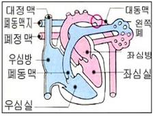
그림 33. 대동맥 축착
◯로 표시한 부분의 대동맥이 축착되어 있다
Copyright ⓒ 2013 John Sangwon Lee, MD., FAAP
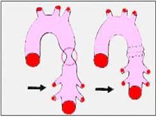
그림 34. 대동맥 수술로 치료하기 전 대동맥 축착(좌)과 수술로 치료한 후 대동맥 축착(우)
◯로 표시한 부분 대동맥이 축착 되어 있다.
Copyright ⓒ 2013 John Sangwon Lee, MD., FAAP
-
대동맥 축착의 크기, 종류, 함께 생겨 있는 동반 선천성 심장혈관 질환, 대동맥 축착 부분의 혈액 흐름의 정도에 따라 증상 징후가 다르다.
-
축착된 대동맥 부분 이하에 있는 대동맥에서 피를 공급받는 신체 부위는 필요 한 만큼 혈 량을 충분히 공급받을 수 없다.
-
따라서 그 신체 부분의 혈색이 창백해질 수 있고 체온이 비정상적으로 낮고 혈압도 낮을 수 있다.
-
그와 반대로, 축착 된 대동맥의 부분이 있는 이전 대동맥 부분에서 피를 공급받는 신체 부분은 필요 이상 혈 량을 더 많이 공급받기 때문에 그 신체 부분의 혈색이 더 붉고 혈압이 더 올라가서 고혈압이 생길 수 있다.
-
대동맥 축착이 대동맥의 어느 부위에 생겨 있는지, 축착 부분의 길이와 정도, 대동맥 축착을 처음 진단 받을 때의 환아 나이, 합병증의 유무 등에 따라 증상 징후가 다르다.
-
경미한 대동맥 축착은 아무 증상 징후가 나타나지 않을 수 있다.
-
그렇지만, 출생 후 어느 때든지 심부전증, 두통, 축착이 되어 있는 대동맥 부분 이전에 있는 대동맥 부분의 혈을 공급받는 상반신에 생긴 고혈압, 축착되어 있는 대동맥 아래에 있는 대동맥의 혈을 공급받는 하반신에 저혈압과 체온 하강, 코피 등의 증상 징후가 생길 수 있다.
-
심한 고혈압으로 두개강 내 뇌혈관이 파열될 수 있다.
-
정상적으로 하반신 혈압이 상반신 혈압보다 조금 더 높은 것이 보통이다.
-
그렇지만 대동맥 축착이 있을 때는 하반신 혈압은 상반신 혈압보다 훨씬 낮을 수 있다.
-
대동맥 축착이 있는 신생아들에게 심장부전증이 생기면서 성장 발육이 지연되고 전신이 쇠약해진다.
-
이 기형을 적기에 수술 치료를 받지 않으면 수명이 20∼40세 정도이다.
대동맥 축착의 진단
-
병력, 증상 징후, 진찰소견 등을 종합하여 이 병이 의심되면 심전도 검사, 가슴 X-선 사진 검사, 심도자 검사, 심초음파 검사, CT 스캔 검사나 그 외 다른 여러 가지 검사로 쉽게 확진할 수 있다.
대동맥 축착의 치료
-
축착된 대동맥 부분의 길이와 정도, 증상 징후, 합병증 유무, 나이 등에 따라 수술치료방법이 다르다.
-
일반적으로 생명에 위협받을 수 있는 증상 징후가 나타나지 않으면, 가능한 한 3∼6세가 될 때까지 기다렸다가 축착된 대동맥 부분을 수술로 제거해 내고 정상적인 양쪽 대동맥 끝 부분을 서로 연결시켜 주는 수술 치료를 한다.
-
축착된 대동맥의 부분이 상당히 길어서 축착된 부분을 제거해 내고 정상 대동맥의 양쪽의 끝 부분을 서로 연결시킬 수 없을 때는 다크론 인공혈관 등으로 연결 수술치료를 할 수 있다.
Coarctation of aorta 대동맥 축착
• The aorta originates from the left ventricle and is the largest of all blood vessels in the human body.
• Total blood volume in the left ventricle passes through the aortic valve at the origin of the aorta and flows into the systemic and coronary arteries.
• A congenital aortic malformation that narrows due to abnormal Coarctation of a part of the aorta is called aortic condensation.
• The portion of the condensed aorta may be very short or longer.
• The degree of condensation may or may not be severe.
• Sometimes a part of the aorta may only be traced. There are several types of Coarctation.
• The most common Coarctation occurs at the origin of the aorta in the thoracic cavity below the left clavicle.
Causes of Coarctation of aorta
• The cause is still unknown.
• Coarctation of aorta is more common in girls with Turner’s syndrome than in normal girls without Turner’s syndrome. • In addition, Coarctation of aorta and other congenital heart anomalies are more likely to occur in children who have congenital malformations in organs of different systems in many branches of the body.
Symptoms signs of Coarctation of aorta
Figure 33. Coarctation of aorta The aorta in the area marked by ◯ is Coarctation. Copyright ⓒ 2013 John Sangwon Lee, MD., FAAP
Figure 34. Coarctation of aorta before treatment with aortic surgery (left) and Coarctation of aorta after treatment with aortic surgery (right) The partial aorta marked with ◯ is condensed. Copyright ⓒ 2013 John Sangwon Lee, MD., FAAP
• Symptoms differ depending on the size and type of aortic condensation, congenital congenital cardiovascular disease, and the degree of blood flow to the area of the Coarctation.
• Parts of the body that receive blood from the aorta below the Coarctation cannot receive enough blood volume as needed.
• Therefore, the color of the body part may become pale, the body temperature may be abnormally low, and the blood pressure may be low.
• Conversely, the part of the body that receives blood from the part of the previous aorta that has a part of the Coarctation receives more blood than is needed, so that part of the body may have redder and higher blood pressure, leading to high blood pressure.
• Symptoms differ depending on where Coarctation occurs in the aorta, the length and extent of the condensation part, the age of the patient at the time of initial diagnosis of Coarctation, and the presence or absence of complications.
• Minor Coarctation may show no symptoms.
• Nevertheless, at any time after birth, heart failure, headache, high blood pressure in the upper body receiving blood from the aortic part before the condensed aorta, hypotension in the lower body receiving blood from the aorta under the condensed aorta. Symptoms such as a drop in body temperature and nosebleeds may occur.
• Severe high blood pressure can cause rupture of blood vessels in the cranial cavity.
• Normally, the lower body blood pressure is slightly higher than the upper body blood pressure.
• However, in the presence of aortic condensation, the lower torso blood pressure can be much lower than the upper torso blood pressure.
• Newborns with Coarctation develop heart failure, delaying growth and debilitating whole body. • If the malformation is not treated in a timely manner, the life expectancy is about 20-40 years old.
Diagnosis of Coarctation of aorta
• If the disease is suspected by taking the medical history, symptoms, signs, examination findings, etc., it can be easily confirmed with an electrocardiogram, chest X-ray, cardiogram, echocardiography, CT scan, or other tests.
Treatment of Coarctation of aorta
• The surgical treatment method differs depending on the length and extent of the condensed aorta, symptoms, signs, complications, and age.
• In general, if there are no signs of life-threatening symptoms, wait until 3-6 years old as possible, then surgically remove the condensed aorta and connect the normal ends of the aorta to each other.
• If the condensed aorta is quite long and the condensed part is removed and the ends of both sides of the normal aorta cannot be connected to each other, connection surgery can be performed with a dacron artificial blood vessel.
출처 및 참조 문헌 Sources and references
- NelsonTextbook of Pediatrics 22ND Ed
- The Harriet Lane Handbook 22ND Ed
- Growth and development of the children
- Red Book 32nd Ed 2021-2024
- Neonatal Resuscitation, American Academy Pediatrics
- www.drleepediatrics.com제11권. 소아청소년 심장 혈관계 질환
- www.drleepediatrics.com제7권 소아청소년 감염병
- Red book 29th-31st edition 2021
- Nelson Text Book of Pediatrics 19th — 21st Edition
- The Johns Hopkins Hospital, The Harriet Lane Handbook, 22nd edition
-
Childhood Emergencies in the Office, Hospital and Community, American Academy of Pediatrics
-
Emergency Medical Service for Children, By Ross Lab. May 1989. p.10
-
Emergency care, Harvey grant, and Robert Murray
-
Emergency Care Transportation of Sick and Injured American Academy of Orthopaedic Surgeons
-
Emergency Pediatrics A Guide to Ambulatory Care, Roger M. Barkin, Peter Rosen
-
Immediate care of the acutely ill and injured, Hugh E. Stephenson, Jr
-
The Critically Ill Child, Diagnosis and Management, Edited by Clement A. Smith
-
Emergency Medical Services for Children: The Role of the Primary Care Provider, America Academy of Pediatrics
-
Quick Reference To Pediatric Emergencies, Delmer J. Pascoe, M.D., Moses Grossman, M.D. with 26 contributors
-
Manual of Emergency Care
-
응급환자관리 정담미디어
-
소아가정간호백과–부모도 반의사가 되어야 한다, 이상원
-
Neonatal Resuscitation American heart Association
-
Neonatology Jeffrey J.Pomerance, C. Joan Richardson
-
Pediatric Resuscitation Pediatric Clinics of North America, Stephen M. Schexnayder, M.D.
-
Pediatric Critical Care, Pediatric Clinics of North America, James P. Orlowski, M.D.
-
Preparation for Birth. Beverly Savage and Dianna Smith
-
Nelson Textbook of Pediatrics 14th ed. Beherman,
-
The Johns Hopkins Hospital, The Harriet Lane Handbook, 18th edition
-
Red book 29th edition 2012
- 소아과학 대한교과서
-
제1권 소아청소년 응급의료 참조문헌과 출처
-
Other
Copyright ⓒ 2015 John Sangwon Lee, MD., FAAP
“부모도 반의사가 되어야 한다”-내용은 여러분들의 의사로부터 얻은 정보와 진료를 대신할 수 없습니다.
“The information contained in this publication should not be used as a substitute for the medical care and advice of your doctor. There may be variations in treatment that your doctor may recommend based on individual facts and circumstances. “Parental education is the best medicine.”