늑막염(흉막염)과 농흉 Pleurisy and Empyema
| 늑막염(흉막염)과 농흉의 개요 및 원인 |
- 폐는 두 겹의 엷은 막으로 둘러싸여 있다.
- 이 두 겹의 엷은 막을 늑막 또는 흉막이라고 한다.
- 폐 표면을 둘러싼 흉막을 폐흉막 또는 폐늑막이라 하고, 흉곽의 내면에 접촉돼있고 폐늑막의 바깥 면에 접해 있는 늑막을 벽흉막이라고 한다.
- 이 두 겹의 늑막(흉막) 사이에 있는 공간을 늑막 강(흉막 강)이라고 한다(그림 111참조).
- 늑막 강 속에는 미끈거리는 체액이 정상적으로 소량 들어있다.
- 결핵균이나 다른 종류의 박테리아, 또는 그 외 다른 병원체에 늑막 강이 1차적으로 감염되면 늑막강 속에 감염병이 생길 수 있다.
- 이렇게 생긴 늑막 강 감염병을 늑막염(Pleurisy/흉막염)이라고 한다.
- 늑막 강에 박테리아 감염이 생기고 그로 인해 늑막 강 속에 감염병이 생기면 늑막 강 감염균의 종류에 따라 짙은 혼탁 고름, 엷은 혼탁 고름, 또는 맑은 혼탁 고름이 늑막 강 속에 괼 수 있다.
- 또 병원체가 감염되지 않았는데 신증후군이나 심장병 등으로 전신의 각 계통의 각 기관과 조직에 체액이 비정상적으로 괼 수 있다.
- 이런 경우, 늑막 강 속에 맑은 체액이 괼 수 있다. 즉 늑막 강 속, 복강 속, 그 외 신체의 다른 계통의 다른 조직과 기관에 체액이 비정상적으로 많이 괼 수 있다.
- 화농성 세균이 늑막 강에 감염되고 그로 인해 늑막 강 감염병이 생기면 늑막 강 속에 고름이 잡힐 수 있다. 이때 늑막 강 속에 짙고 혼탁한 고름이 괸 늑막염이 생길 수 있다.
- 이런 늑막염을 농흉(Empyema)이라고 한다.
- 폐렴을 앓는 소아청소년 150명 중 1명에게 농흉이 생길 수 있고 소아 청소년 입원 환아 1000명 증 0.4~6명에게 농흉이 있었다.
- 최근에는 소아 청소년들에게 농흉이 더 자주 발견되고, 특히 폐렴연쇄상구균 폐렴이 소아 청소년들에게 있을 때 가슴 초음파 검에 8~14%에서 농흉이 발견된다고 한다(출처: Pediatric News, March 2004).
- 박테리아, 바이러스, 그 밖의 다른 병원체가 늑막염 및, 또는 농흉을 일으킬 수 있다.
- 황색 포도상구균, A군 베타 용혈성 연쇄상구균, B형 헤모필러스 인플루엔자균, 폐렴연쇄상구균, 그밖에 다른 종류의 박테리아가 늑막강 속에 직접 또는 간접적으로 감염되어 늑막염(흉막염)이나 농흉을 일으킬 수 있다.
- 폐렴을 일으킨 박테리아가 늑막강 속으로 1차적으로 감염되어 늑막염이나 농흉을 일으킬 수 있다.
- 결핵균, 바이러스, 마이코플라스마 등 병원체가 직접 또는 간접적으로 늑막강 속이나 폐에 일차적으로 감염 되어 늑막염을 일으킬 수 있다. 그런 늑막염으로 생긴 늑막강 속 체액은 맑고 투명한 것이 보통이다.
- 췌장염, 요독증, 신증후군, 심장병, 간경변증, 악성 종양, 백혈병 등을 앓을 때 능막강 속에 맑고 투명한 체액이 2차적으로 괼 수 있다.
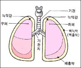
그림 111. 늑막, 늑막강과 폐.
Copyright ⓒ 2012 John Sangwon Lee, MD., FAAP
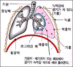
그림 112. 긴장성 기흉.
Copyright ⓒ 2012 John Sangwon Lee, MD., FAAP
| 늑막염(흉막염)과 농흉의 증상 징후 |
- 늑막염이나 농흉을 일으킨 원인, 다른 동반 병의 유무, 합병증의 유무, 병의 중증도에 따라 증상 징후가 다르다.
- 박테리아 감염으로 늑막염이 생겼을 때는 늑막염이 생긴 쪽의 가슴이 아프고 열이 나며 호흡곤란이 생긴다.
- 늑막염과 폐렴이 동시에 생길 수 있다. 이때 두 가지 병으로 생기는 증상 징후가 함께 나타날 수 있다.
출처 및 참조문헌
- 신생아 흡인 폐렴,
- 황색 포도상구균 폐렴,
- 기흉 참조
| 늑막염(흉막염)과 농흉의 진단 |
- 증상 징후, 진찰소견, 가슴 X-선 사진, 피 검사와 소변 검사, 세균 배양검사 등을 종합하여 진단할 수 있다.
- 늑막염이 결핵균 감염으로 생겼을 때는 결핵 피부 반응검사 결과가 양성으로 나타난다.
- 원인을 더 확실하게 알아보기 위해 늑막 강 내 체액이나 고름을 빼고 그 고름으로 그람 염색 현미경 세균 검사, 세균 배양검사 및 다른 여러 가지 검사로 진단할 수 있다.
| 늑막염(흉막염)과 농흉의 치료 |
- 늑막염을 일으킨 원인과 합병증의 유무 등에 따라 치료한다.
- 결핵균에 의한 늑막염이 있을 때는 결핵 치료약으로 치료한다.
- 또 농흉의 경과를 3기로 나눌 수 있다.
- 각기에 따라 치료를 달리할 수 있다.
- 황색 포도상구균 감염이나 다른 종류의 박테리아 감염으로 생긴 농흉이 있을 때는 늑막강에 있는 고름을 흉막 튜브(관)를 통한 배농 치료 하거나
- 흉막천자 수술 배농 치료,
섬유소 융해제 치료, - 또는 외과적 수술 배농 치료 방법과 동시 적절한 항생제 정맥주사로 치료할 수 있다.
- 신증후군이나 심장병 등에 의해 늑막강 속에 늑막 액이 괼 때는 신증후군이나 심장병 등 원발성 원인이 되는 병을 치료해 주면 늑막강 속에 괴었던 체액이 자연히 빠져 없어진다.
- 늑막강 내 Urokinase 치료 또는 비디오를 통한 흉막강 내시경 외과적 치료로 치료 할 수 있다. (출처: Pediatric News, March 2004)
Pleurisy and Empyema 늑막염(흉막염)과 농흉
Overview and causes of pleurisy (pleurisy) and empyema
• The lungs are surrounded by two layers of thin film.
• These two layers of thin film are called the pleura or pleura.
• The pleura surrounding the lung surface is called the pulmonary pleura or pulmonary pleura, and the pleura that is in contact with the inner surface of the thorax and the outer surface of the lung pleura is called the parietal pleura.
• The space between these two layers of the pleura (pleura) is called the pleural cavity (pleural cavity) (see Figure 111).
• The pleural cavity normally contains a small amount of slimy body fluid.
• Primary infection of the pleural cavity with Mycobacterium tuberculosis, other types of bacteria, or other pathogens can lead to infectious diseases in the pleural cavity.
• The resulting pleural cavity infection is called pleurisy.
• If a bacterial infection occurs in the pleural cavity and the resulting infectious disease occurs in the pleural cavity, depending on the type of pleural cavity infectious bacteria, thick cloudy pus, light cloudy pus, or clear cloudy pus may accumulate in the pleural cavity. • In addition, even though the pathogen has not been infected, body fluids may bleed abnormally into organs and tissues in each system of the body due to nephrotic syndrome or heart disease.
• In this case, clear body fluid may collect in the pleural cavity. In other words, abnormally large amounts of fluid may be collected in the pleural cavity, the abdominal cavity, and other tissues and organs of other systems of the body.
• If purulent bacteria infect the pleural cavity, resulting in a pleural cavity infection, pus can be trapped in the pleural cavity. At this time, pleurisy may develop in the pleural cavity with dark and cloudy pus. • This pleurisy is called empyema.
• One in 150 children and adolescents with pneumonia may develop empyema, and 0.4 to 6 children in 1,000 hospitalized children and adolescents had empyema.
• In recent years, empyema is found more frequently in children and adolescents, especially when pneumonia streptococcal pneumonia is present in children and adolescents.
• Bacteria, viruses, and other pathogens can cause pleurisy and/or empyema.
• Staphylococcus aureus, group A beta hemolytic streptococcus, Haemophilus influenzae type B, streptococci pneumonia, and other types of bacteria can directly or indirectly infect the pleural cavity and cause pleurisy (pleurisy) or empyema.
• The bacteria that cause pneumonia can become infected primarily into the pleural cavity, causing pleurisy or empyema.
• Pathogens such as tuberculosis bacteria, viruses, and mycoplasma may directly or indirectly infect the pleural cavity or lungs and cause pleurisy. The fluid in the pleural cavity caused by such pleurisy is usually clear and transparent.
• When suffering from pancreatitis, uremia, nephrotic syndrome, heart disease, cirrhosis, malignant tumors, leukemia, etc., clear and transparent body fluids may be collected in the neural cavity secondarily.
Figure 111. Pleural, pleural cavity and lungs. Copyright ⓒ 2012 John Sangwon Lee, MD., FAAP
Figure 112. Tension pneumothorax. Copyright ⓒ 2012 John Sangwon Lee, MD., FAAP
Symptoms Signs of pleurisy (pleurisy) and empyema
• Symptoms and signs differ depending on the cause of pleurisy or empyema, the presence of other comorbidities, complications, and severity of the disease.
• When pleurisy occurs due to a bacterial infection, the chest on the side where the pleurisy occurs is painful, fever, and shortness of breath.
• Pleurisy and pneumonia can develop at the same time. At this time, symptoms of the two diseases may appear together. Sources and references
• neonatal aspiration pneumonia,
• Staphylococcal pneumonia,
• See pneumothorax
Diagnosis of pleurisy (pleurisy) and empyema
• Symptom signs, examination findings, chest X-rays, blood and urine tests, and bacterial culture tests can be comprehensively diagnosed. • When pleurisy is caused by a Mycobacterium tuberculosis infection, the tuberculosis skin test result is positive. • To further determine the cause, fluid or pus in the pleural cavity can be drained and diagnosed with Gram-stained microscopic bacterial tests, bacterial culture tests, and several other tests.
Treatment of pleurisy (pleurisy) and empyema
• Treat according to the cause of the pleurisy and the presence or absence of complications.
• If you have pleurisy caused by Mycobacterium tuberculosis, treat with tuberculosis medication. • Also, the course of empyema can be divided into three stages.
• Treatment can be different for each.
• If you have an empyema caused by a Staphylococcus aureus infection or another type of bacterial infection, the pus in the pleural cavity is drained through a pleural tube (tube) or
• pleural puncture surgery drainage treatment, Fibrinolytic therapy,
• Or, it can be treated with an appropriate intravenous injection of antibiotics at the same time as surgical drainage treatment.
• When the pleural fluid is squeezed in the pleural cavity due to nephrotic syndrome or heart disease, treatment of the primary cause such as nephrotic syndrome or heart disease will cause the body fluid accumulated in the pleural cavity to disappear naturally.
• It can be treated with intrapleural Urokinase treatment or pleural endoscopic surgery through video. (Source: Pediatric News, March 2004)
출처 및 참조 문헌 Sources and references
- NelsonTextbook of Pediatrics 22ND Ed
- The Harriet Lane Handbook 22ND Ed
- Growth and development of the children
- Red Book 32nd Ed 2021-2024
- Neonatal Resuscitation, American Academy Pediatrics
- www.drleepediatrics.com제7권 소아청소년 감염병
-
www.drleepediatrics.com제8권 소아청소년 호흡기 질환
-
Red book 29th-31st edition 2021
- Nelson Text Book of Pediatrics 19th — 21st Edition
-
The Johns Hopkins Hospital, The Harriet Lane Handbook, 22nd edition
-
Childhood Emergencies in the Office, Hospital and Community, American Academy of Pediatrics
-
Emergency Medical Service for Children, By Ross Lab. May 1989. p.10
-
Emergency care, harvey grant and robert murray
-
Emergency Care Transportation of Sick and Injured American Academy of Orthopaedic Surgeons
-
Emergency Pediatrics A Guide to Ambulatory Care, Roger M. Barkin, Peter Rosen
-
Immediate care of the acutely ill and injured, Hugh E. Stephenson, Jr
-
The Critically Ill Child, Diagnosis and Management, Edited by Clement A. Smith
-
Emergency Medical Services for Children: The Role of the Primary Care Provider, America Academy of Pediatrics
-
Quick Reference To Pediatric Emergencies , Delmer J. Pascoe, M.D., Moses Grossman, M.D. with 26 contributors
-
Manual of Emergency Care
-
응급환자관리 정담미디어
-
소아가정간호백과–부모도 반의사가 되어야 한다, 이상원
-
Neonatal Resuscitation American heart Association
-
Neonatology Jeffrey J.Pomerance, C. Joan Richardson
-
Pediatric Resuscitation Pediatric Clinics of North America, Stephen M. Schexnayder, M.D.
-
Pediatric Critical Care, Pediatric Clinics of North America, James P. Orlowski, M.D.
-
Preparation for Birth. Beverly Savage and Dianna Smith
-
Nelson Textbook of Pediatrics 14th ed. Beherman,
-
The Johns Hopkins Hospital, The Harriet Lane Handbook, 18th edition
-
Red book 29th edition 2012
-
소아과학 대한교과서
-
제1권 소아청소년 응급의료 참조문헌과 출처
-
Other
- 신생아 폐렴 Neonatal pneumonia(Pneumonia of newborn/pneumonia in newborn)
Copyright ⓒ 2014 John Sangwon Lee, MD., FAAP
“부모도 반의사가 되어야 한다”-본 사이트의 내용은 여러분들의 의사로부터 얻은 정보와 진료를 대신할 수 없습니다.
“The information contained in this publication should not be used as a substitute for the medical care and advice of your doctor. There may be variations in treatment that your doctor may recommend based on individual facts and circumstances.
“Parental education is the best medicine.“