기흉 Pneumothorax
기흉의 개요와 원인
- 늑막강 속에 공기가 차 있는 병을 기흉이라고 한다.
- 갓 태어난 신생아에게 생기는 기흉의 원인([부모도 반의사가 되어야 한다-소아가정간호백과]-제6권 신생아 성장 발육 양호 질병-기흉 참조)과 신생아기 이후 아이에게 생기는 기흉의 원인이 다를 수 있다(그림 112 기흉 참조).
- 확실한 원인 없이 생기는 기흉을 1차성 자연 기흉(Primary spontaneous pneumothorax), 이미 존재한 질병과 관련되어 생긴 기흉을 2차성 기흉(Secodary spontaneous pneumothorax)이라고 한다.
- 1차성 자연 기흉은 남성 만 명 중 7.4명, 여성 만 명 중 1.2명에게 발생한다.
- 체질적으로 외배엽 체형을 가진 사람들에게 좀 더 생길 수 있다.
- 1차성 자연 기흉이 있었던 가족 병력이 있는 사람들에게 더 잘 생긴다(참조문헌Contemporary pediatrics, December, 2008. p.504).
- 황색 포도상구균 폐렴(폐렴 참조)·기관지 천식·폐결핵, 만성 차단성 폐질환, 에이즈, 뉴모 시스티스 폐렴, 그밖에 다른 여러 종류의 폐렴·폐농양·폐낭포·폐기흉·흉곽수술·흉곽 외상 등으로 2차성 기흉이 생길 수 있다.
- 즉 다른 병이 이미 있고 그 병으로 인해 생긴 기흉을 2차성 기흉이라고 한다.
- 아무런 이유도 없이 기흉이 생길 수 있다. 이런 기흉을 1차성(원발성) 자연 기흉이라고 한다.
- 참고로, 늑막강 내 압은 음압인 것이 정상이고, 기관지 내 압이나 폐기포 내 압은 양압인 것이 정상적이다.
- 기흉이 있는 늑막강 내 압이 기관지 내 압이나 폐기포 내 압보다 더 높을 때가 있다.
- 기흉의 압이 비정상적으로 상당히 높고 기흉이 상당히 커질 때는 기흉이 있는 쪽의 폐가 기흉으로 눌려서 쪼그라질 수 있다.
- 이런 기흉을 긴장성 기흉(Tension pneumothorax)라고 한다(그림 113 참조).
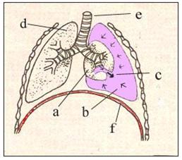
그림113.늑막강 속에 공기가 차 있는 기흉
a-쪼그라진 폐, b-기흉, c-세기관지나 폐포 등에서 공기가 늑막강 속으로 새 들어간다, d-정상 우폐, e-기관, f-횡격막.
출처-Copyrightⓒ 2001 John Sangwon Lee,MD., FAAP
기흉의 증상 징후
- 기흉의 종류와 중증도, 원인, 합병증 유무 등에 따라 증상 징후가 다르다.
- 기흉이 흡인 폐렴(신생아 흡인성 폐렴 참조)이나 황색 포도상구균 폐렴, 또는 기관지 천식 등이 있을 때 생긴 2차성 기흉의 증상 징후는 원래 있던 병으로 생긴 증상 징후와 기흉으로 생긴 증상 징후와 함께 나타나는 것이 보통이다.
- 긴장성 기흉이 있을 때는 갑자기 호흡곤란이 생길 수 있고, 안색이 창백하면서 안절부절 못하고 기흉이 생긴 쪽 가슴이 아플 수 있다.
- 기흉이 경미할 때는 아무런 증상 징후가 없을 수 있다.
- 1차성 자연 기흉이 심할 때는 기흉이 있는 쪽의 가슴이 갑자기 아프고 숨이 가쁘고 빈맥이 생기고 숨소리가 감소되고 기침을 할 수 있다.
기흉의 진단
- 병력·증상 징후·진찰소견 등을 종합해서 이 병이 의심되면, 가슴 X-선 사진 검사로 진단할 수 있다.
- 기흉을 일으킨 원인에 따라 진단 방법이 다르다.
기흉의 치료
- 기흉의 종류, 중증도, 기흉을 일으킨 원인에 따라 치료가 다르다.
- 1차성 자연 기흉의 일부는 약 1~2주일 동안 육체적 안정을 취하면 자연히 치료될 수 있다.
- 주사바늘로 늑막강 내 공기를 빼주고 관찰하면 자연적으로 회복될 수 있다.
- 긴장성 기흉이 심할 때는 생명에도 위험할 수 있다.
- 늑막강이 있는 공기를 흉벽 관(Chest tubes)으로 빼주고 쪼그라진 폐를 정상적으로 회복시키기는 치료를 해준다.
- 때로는 작은 흉벽 관이나 큰 주사 바늘을 기흉이 있는 늑막강 속에 넣고 공기를 응급으로 빼내는 치료도 한다.
- 기흉이 있는 늑막강 속에 넣었던 흉벽 관은 공기가 더 이상 나오지 않을 때까지 계속 그 늑막강 속에 삽입 해 놓았다가 공기가 더 이상 나오지 않으면 빼내는 식으로 치료한다.
- 이런 치료 방법으로 완치가 안 되면 늑막강 속으로 공기가 새어나오게 하는 폐의 부분이나 세기관지 부분을 수술로 꿰매어 치료하기도 한다.
- 그리고 기흉을 일으킨 원인을 동시 치료해 준다.
Pneumothorax
Overview and Causes of Pneumothorax
• A disease in which air is filled in the pleural cavity is called pneumothorax.
• The causes of pneumothorax in newborn babies ([Parents should also become at-lerast half doctors – Encyclopedia of Pediatric Home Nursing] – Volume 6 Diseases with Good Growth and Development in Newborns – Pneumothorax) may be different from the causes of pneumothorax in children after the neonatal period. (See Figure 112 Pneumothorax).
• A pneumothorax that occurs without a definite cause is called primary spontaneous pneumothorax, and a pneumothorax associated with an existing disease is called secondary spontaneous pneumothorax.
• Primary spontaneous pneumothorax occurs in 7.4 out of 10,000 men and 1.2 out of 10,000 women.
• It is more likely to occur in people with an ectodermal body type.
• More likely to occur in people with a family history of primary spontaneous pneumothorax (Contemporary pediatrics, December, 2008. p.504).
• Staphylococcus aureus pneumonia (see Pneumonia) • Bronchial asthma • Pulmonary tuberculosis, chronic obstructive pulmonary disease, AIDS, pneumocystis pneumonia, other types of pneumonia
• Pulmonary abscess
• Pneumococcal cysts
• Pneumothorax
• Thoracic surgery
• Thoracic trauma, etc. can cause secondary pneumothorax.
• In other words, a pneumothorax caused by another disease is called secondary pneumothorax.
• Pneumothorax can occur for no reason. Such pneumothorax is called primary (primary) spontaneous pneumothorax.
• For reference, it is normal for the intrapleural pressure to be negative, and it is normal for the intrabronchial or alveolar pressure to be positive.
• In pneumothorax, the intrapleural pressure is sometimes higher than the intrabronchial or alveolar pressure.
• When the pressure of the pneumothorax is abnormally high and the pneumothorax becomes quite large, the lung on the side of the pneumothorax may be compressed by the pneumothorax and contracted.
• This pneumothorax is called a tension pneumothorax (see Figure 113).
Figure 113. Pneumothorax with air in the pleural space a – shriveled lung, b – pneumothorax, c – leak of air into the pleural space from bronchioles or alveoli, d – normal right lung, e – trachea, f – diaphragm. Source-Copyrightⓒ 2001 John Sangwon Lee, MD., FAAP
Symptoms, signs of Pneumothorax
• Symptoms differ depending on the type and severity of pneumothorax, cause, and presence or absence of complications.
• Symptoms, signs of secondary pneumothorax caused when pneumothorax is aspiration pneumonia (see neonatal aspiration pneumonia), staphylococcal pneumonia, or bronchial asthma should be accompanied by symptomatic signs of the original disease and symptoms of pneumothorax. is average.
• When there is tension pneumothorax, shortness of breath may occur suddenly, the complexion may be pale and restless, and the chest on the side of the pneumothorax may be painful.
• When pneumothorax is mild, there may be no symptoms.
• When primary spontaneous pneumothorax is severe, there is sudden chest pain on the side of the pneumothorax, shortness of breath, tachycardia, decreased breathing sound, and coughing.
Diagnosis of pneumothorax
• If the disease is suspected based on the medical history, symptom signs, and examination findings, it can be diagnosed by chest X-ray examination.
• Diagnosis methods differ depending on the cause of pneumothorax.
Treatment of pneumothorax
• Treatment differs depending on the type, severity, and cause of the pneumothorax.
• Some of the primary spontaneous pneumothorax can be cured spontaneously with physical rest for about 1 to 2 weeks.
• The patient can recover naturally by evacuating the pleural cavity with a needle and observing it.
• Severe tension pneumothorax can be life-threatening.
• Treatment is to remove the air from the pleural cavity through the chest tubes and restore the shriveled lungs to normal.
• Sometimes, a small chest wall tube or a large needle is placed into the pleural space where the pneumothorax is located and air is expelled as an emergency.
• The chest wall tube inserted into the pleural cavity with pneumothorax is continuously inserted into the pleural cavity until no more air comes out.
• If there is no cure with this treatment method, the part of the lung or bronchioles that allows air to leak into the pleural space may be surgically stitched.
• It also treats the cause of pneumothorax at the same time.
출처 및 참조 문헌 Sources and references
- NelsonTextbook of Pediatrics 22ND Ed
- The Harriet Lane Handbook 22ND Ed
- Growth and development of the children
- Red Book 32nd Ed 2021-2024
- Neonatal Resuscitation, American Academy Pediatrics
- www.drleepediatrics.com제6권 신생아 성장 발육 육아 질병
- www.drleepediatrics.com제7권 소아청소년 감염병
-
www.drleepediatrics.com제8권 소아청소년 호흡기 질환
-
Red book 29th-31st edition 2021
- Nelson Text Book of Pediatrics 19th — 21st Edition
-
The Johns Hopkins Hospital, The Harriet Lane Handbook, 22nd edition
-
Childhood Emergencies in the Office, Hospital and Community, American Academy of Pediatrics
-
Emergency Medical Service for Children, By Ross Lab. May 1989. p.10
-
Emergency care, Harvey grant, and Robert Murray
-
Emergency Care Transportation of Sick and Injured American Academy of Orthopaedic Surgeons
-
Emergency Pediatrics A Guide to Ambulatory Care, Roger M. Barkin, Peter Rosen
-
Immediate care of the acutely ill and injured, Hugh E. Stephenson, Jr
-
The Critically Ill Child, Diagnosis and Management, Edited by Clement A. Smith
-
Emergency Medical Services for Children: The Role of the Primary Care Provider, America Academy of Pediatrics
-
Quick Reference To Pediatric Emergencies, Delmer J. Pascoe, M.D., Moses Grossman, M.D. with 26 contributors
-
Manual of Emergency Care
-
응급환자관리 정담미디어
-
소아가정간호백과–부모도 반의사가 되어야 한다, 이상원
-
Neonatal Resuscitation American heart Association
-
Neonatology Jeffrey J.Pomerance, C. Joan Richardson
-
Pediatric Resuscitation Pediatric Clinics of North America, Stephen M. Schexnayder, M.D.
-
Pediatric Critical Care, Pediatric Clinics of North America, James P. Orlowski, M.D.
-
Preparation for Birth. Beverly Savage and Dianna Smith
- Infectious disease of children, Saul Krugman, Samuel L Katz, Ann A. Gershon, Catherine Wilfert
-
The Harriet Lane Handbook 19th Edition
-
소아과학 대한교과서
-
제1권 소아청소년 응급의료 참조문헌과 출처
-
Other
Copyright ⓒ 2015 John Sangwon Lee, MD., FAAP
“부모도 반의사가 되어야 한다”-내용은 여러분들의 의사로부터 얻은 정보와 진료를 대신할 수 없습니다.
“The information contained in this publication should not be used as a substitute for the medical care and advice of your doctor. There may be variations in treatment that your doctor may recommend based on individual facts and circumstances. “Parental education is the best medicine.”