골절 Fractures
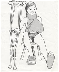
그림 175. 정형외과 환자
Copyright ⓒ 2011 John Sangwon Lee, M.D., FAAP
- 숱하게 많은 종류의 골절과 골절의 증상 진단 치료에 관하여 이 책에서 다 설명할 수 없다.
- 나이에 따라, 골절의 정도에 따라, 골절된 골의 종류와 부위에 따라, 또는 다른 여러 가지 조건에 따라 골절의 원인, 증상 징후, 진단 치료가 다를 수 있다.
- 관찰적 치료, 슬링치료, 목발치료, 부목치료, 기브스치료, 수술치료, 핀치료, 몇가지 배합치료 등으로 골절을 치료할 수 있다.
그림과 X선 사진으로 보는 정상 골격과 골절, 탈구
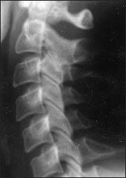
사진 176. 경부 척주 측면 X선 사진.
경부 척주는 7개 척추뼈(Cervical Vertebra/경추)로 이루어져 있다. 한 개 경추나 여러 개 경추가 안전사고 등으로 골절될 수 있다.
Copyright ⓒ 2011 John Sangwon Lee, M.D., FAAP
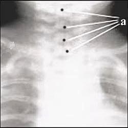
사진 177. 경부 척주 전면 X선 사진
a-경부 척주에 있는 경추, 경부 척주는 7개 척추로 이루어졌다.
Copyright ⓒ 2011 John Sangwon Lee, M.D., FAAP
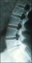
사진 178. 요부 척주 측면 X선 사진
요부 척주는 총 5개 척추뼈(요추)로 이루어졌다.
Copyright ⓒ 2011 John Sangwon Lee, M.D., FAAP
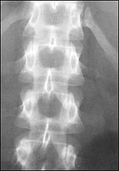
사진 179. 흉부 척주와 요부 척주의 전면 X선 사진.
흉부 척주는 총 12개 척추뼈((흉추)로 이루어졌다.
Copyright ⓒ 2011 John Sangwon Lee, M.D., FAAP
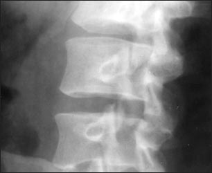
사진 185. 요부 척주의 측면 X선 사진
요부 척주는 총 5개 척추뼈(요추)로 이루어졌다.
Copyright ⓒ 2011 John Sangwon Lee, M.D., FAAP
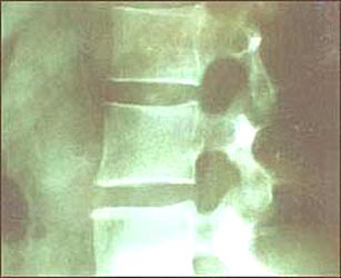
사진 186. 요부 척주의 측면 X선 사진
Copyright ⓒ 2011 John Sangwon Lee, M.D., FAAP
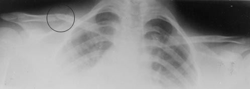
사진 187. ◯내 우 쇄골 골절과 좌 정상 쇄골
Copyright ⓒ 2011 John Sangwon Lee, M.D., FAAP
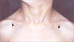
사진 188. 좌우 횡으로 두드러진 부위에 좌우 쇄골이 있다.
Copyright ⓒ 2011 John Sangwon Lee, M.D., FAAP
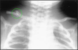
사진 189. 우 쇄골 골절
◯으로 표시된 쇄골 부위가 골절됐다.
Copyright ⓒ 2011 John Sangwon Lee, M.D., FAAP
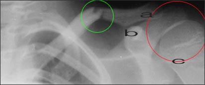
사진 190. ◯내 좌 쇄골 골절
어깨 관절이 ◯ 내에 있다.
Copyright ⓒ 2011 John Sangwon Lee, M.D., FAAP
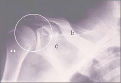
사진 193. 우 어깨관절 X 선 사진
우 어깨관절이 ◯내에 있다.
a-상완골, b-쇄골, c-견갑골
Copyright ⓒ 2011 John Sangwon Lee, M.D., FAAP
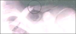
사진 194. ◯내 좌 쇄골 골절
Copyright ⓒ 2011 John Sangwon Lee, M.D., FAAP
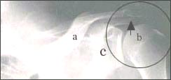
사진 195. ◯어깨관절과 쇄골 골절
↑로 가르킨 부위 쇄골이 골절됐다.
a-쇄골, b-상완골, c-견갑골
Copyright ⓒ 2011 John Sangwon Lee, M.D., FAAP
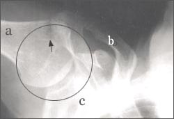
사진 196. ◯내 어깨관절(견갑관절)
↑로 가리킨 부위 상완골 두부가 골절됐다.
a-상완골, b-쇄골, c-견갑골
Copyright ⓒ 2011 John Sangwon Lee, M.D., FAAP
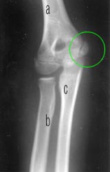
사진 197. 팔꿈치 관절(주관절)의 전면 X선 사진
a-상완골,b-요골, c-척골, ◌ 내에 상완골 내측와가 골절됐다.
Copyright ⓒ 2011 John Sangwon Lee, M.D., FAAP
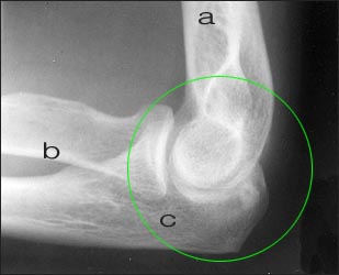
사진 198. 정상 팔꿈치 관절(주관절)(◌ 내) 측면 X선 사진
a-상완골, b-요골, b-척골
Copyright ⓒ 2011 John Sangwon Lee, M.D., FAAP
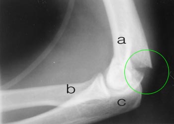
사진 199. ◯내 상완골 골절
a-상완골, b-요골, c-척골
Copyright ⓒ 2011 John Sangwon Lee, M.D., FAAP
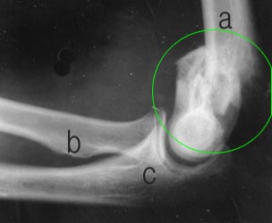
사진 200. ◯ 내 심한 상완골 분쇄 골절
a-상완골, b-요골, c-척골
Copyright ⓒ 2011 John Sangwon Lee, M.D., FAAP
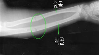
사진 201. ◌내 요골 골절과 척골 골절
Copyright ⓒ 2011 John Sangwon Lee, M.D., FAAP
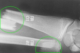
사진 202. 큰 ◌내 요골 골절과 작은 ◌ 내에 척골 골절
Copyright ⓒ 2011 John Sangwon Lee, M.D., FAAP
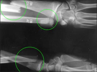
사진 203. 요골 골절과 척골 골절(상,하)
Copyright ⓒ 2011 John Sangwon Lee, M.D.,FAAP
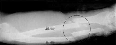
사진 204. ◌내 요골 골절과 척골 골절
Copyright ⓒ 2011 John Sangwon Lee, M.D.. FAAP
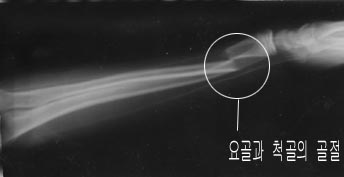
사진 205. ◌내 요골 골절 척골 골절
Copyright ⓒ 2011 John Sangwon Lee, M.D.. FAAP
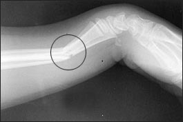
사진 206. ◌내 요골과 척골이 골절됐다.
Copyright ⓒ 2011 John Sangwon Lee, M.D.. FAAP
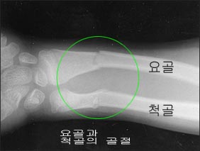
사진 207. ◌내 요골 골절과 척골 골절
Copyright ⓒ 2011 John Sangwon Lee, M.D.. FAAP
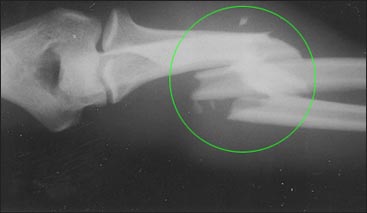
사진 208. 0내 요골과 척골의 복합 골절
Copyright ⓒ 2011 John Sangwon Lee, M.D.. FAAP
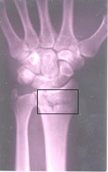
사진 209. □내 요골 골절.
Copyright ⓒ 2011 John Sangwon Lee, M.D.. FAAP
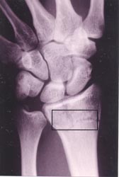
사진 210. □내 요골 골절.
Copyright ⓒ 2011 John Sangwon Lee, M.D.. FAAP
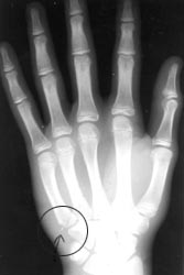
사진 211. ◯내 손뼈 골절
한쪽 손에 14개 수지골(손가락뼈)이 있고, 8개 수근골이 있다.
각 손가락에 해당하는1~5 중수골이 있다. 그 중 한개 뼈나 여러 개 손뼈가 골절될 수 있다. 손뼈의 골단, 골간, 성장판 등에 골절이 생길 수 있다.
Copyright ⓒ 2011 John Sangwon Lee, M.D.. FAAP
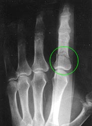
사진 212. ◯내 둘째 손가락 뼈의 첫 째 수지골 골절.
Copyright ⓒ 2011 John Sangwon Lee, M.D.. FAAP
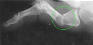
사진 213. ◯내 엄지손가락 중수골 골절
Copyright ⓒ 2011 John Sangwon Lee, M.D.. FAAP

사진 214. ◌내 엄지손가락 첫째 수지골 골절
Copyright ⓒ 2011 John Sangwon Lee, M.D.. FAAP
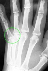
사진 215. ◌내 새끼손가락 중수골과 새끼손가락 첫째 수지골 사이에 있는 관절이 탈구 되어있고 골절도 생겼다.
Copyright ⓒ 2011 John Sangwon Lee, M.D.. FAAP
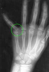
사진 216. ◌내 새끼손가락 중수골과 새끼손가락 수지골 사이 관절이 골절 됐고 탈구도 생겼다.
Copyright ⓒ 2011 John Sangwon Lee, M.D.. FAAP
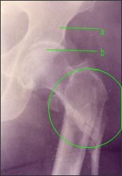
사진 217. 대퇴골 골절(◌ 내)
a-좌골, b-대퇴골 두부
Copyright ⓒ 2011 John Sangwon Lee, M.D.. FAAP
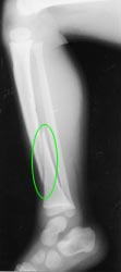
사진 218. 경골 골절(◌내)
Copyright ⓒ 2011 John Sangwon Lee, M.D.. FAAP
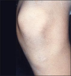
사진 219. 무릎 관절(슬관절)
Copyright ⓒ 2011 John Sangwon Lee, M.D.. FAAP
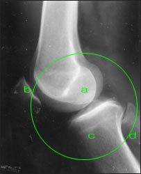
사진 220. ○내 정상 무릎 관절(슬관절)
a-대퇴골 하단, b-슬개골, c-경골, d-비골
Copyright ⓒ 2011 John Sangwon Lee, M.D.. FAAP
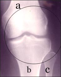
사진 221. 무릎 관절 전후 면 X선 사진
a-대퇴골 하단, b-경골, c-비골
◌내 무릎관절(슬관절).
Copyright ⓒ 2011 John Sangwon Lee, M.D.. FAAP
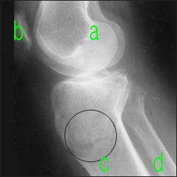
사진 222. 무릎 관절의 측면 X선 사진
a-대퇴골 하단, b-슬개골, c-경골, d-비골
◌내 경골 골절.
Copyright ⓒ 2011 John Sangwon Lee, M.D.. FAAP
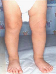
사진 223. 유아의 정상 다리와 발
Copyright ⓒ 2011 John Sangwon Lee, M.D.. FAAP
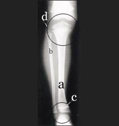
사진 224. 무릎 관절 전후면 X선 사진
a-경골, b-비골, c◯-발목 관절, d◯-무릎 관절
Copyright ⓒ 2011 John Sangwon Lee, M.D.. FAAP
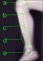
사진 226.◌내 경골 골절
a◯-무릎 관절, b-경골, c-비골, d◯-경골 골절, e◯-발목 관절
Copyright ⓒ 2011 John Sangwon Lee, M.D.. FAAP
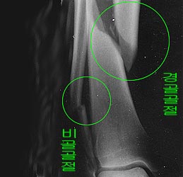
사진 227. 큰 ◌내 경골 골절, 작은 ◌내 비골의 골절
Copyright ⓒ 2011 John Sangwon Lee, M.D.. FAAP
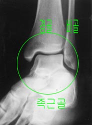
사진 228. 정상 발목(◯내)과 발 전 면 X선 사진
Copyright ⓒ 2011 John Sangwon Lee, M.D.. FAAP
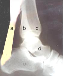
사진 229. 정상 발목과 아킬레스 건, 발목 측면 X선 사진
a-아킬레스건, b-비골 , c-경골, d-거골, e- 종골
Copyright ⓒ 2011 John Sangwon Lee, M.D.. FAAP
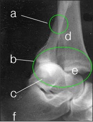
사진 230. 심한 발목 관절 골절과 탈구
a-비골 골절, b◯-발목 관절, c-거골, d-경골, e-경골 두부 탈구, f-종골
Copyright ⓒ 2011 John Sangwon Lee, M.D.. FAAP
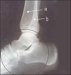
사진231. 발목관절 뼈와 족근골 측면 X선 사진(◌내)
a-경골, b-비골
한쪽 발에 7개 족근골(거골/Talus bone, 종골/Calcaneus bone, 3개 설상골/Three cuneiform bones, 주사위뼈/ Cuboid bone , 주상골/Navicular bone)이 있다. 참고로 한쪽 손에 8개의 수근골이 있다. 족근골과 비골, 경골이 함께 발목관절을 이룬다. 족근골들은 위로 경골과 비골 의 하위부분에 연결되고 아래로 중족골에 연결된다. 족근골이 골절될 수도 있다.
Copyright ⓒ 2011 John Sangwon Lee, M.D.. FAAP
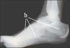
사진 232. 발 측면 X선 사진.
b-발뼈는 족근골, 중족골, 족지골으로 이루어진다.
Copyright ⓒ 2011 John Sangwon Lee, M.D.. FAAP
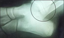
사진 234. 발목 관절 골절과 탈구
◯내 심한 경골 비골 골절과 탈구.
Copyright ⓒ 2011 John Sangwon Lee, M.D.. FAAP
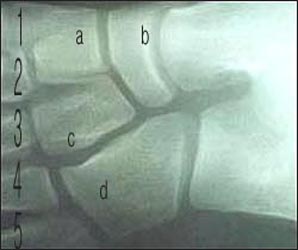
사진 235. 중족골(1, 2, 3, 4, 5)
발 중부에 있는 발뼈들을 중족골이라 한다.
한쪽 발에 5개 중족골이 있고 이 중족골은 뒤로 족근골에 연결되고 앞으로 지절골(발가락뼈)에 연결된다.
a-둘째 쐐기골(둘째 설상골), b-주상골, c- 셋째 쐐기골(셋째 설상골), d- 입방골
Copyright ⓒ 2011 John Sangwon Lee, M.D.. FAAP
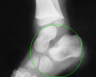
사진 236. 발 후부에 있는 족근골(◌ 내)
Copyright ⓒ 2011 John Sangwon Lee, M.D.. FAAP
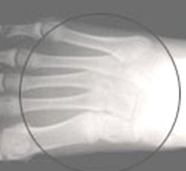
사진 237. ◌내 중족골(1~5)과 족근골
발 중부(발허리)에 있는 발뼈들을 중족골이라 한다.
한쪽 발에 5개 중족골이 있다.
중족골 골절이 생길 수 있다.
Copyright ⓒ 2011 John Sangwon Lee, M.D.. FAAP
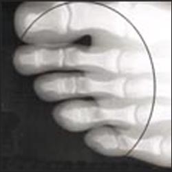
사진 238.◯내 지절골(발가락뼈)
발가락에 있는 발가락뼈를 지절골이라 한다.
14개 지절골이 있다.
지절골에 골절이 생길 수 있다. 엄지 발가락을 무지라고 한다.
Copyright ⓒ 2011 John Sangwon Lee, M.D.. FAAP
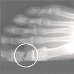
사진 239. ◯내 무지 첫째 지절골 골절(발가락뼈 골절)
한쪽 발에 총 27개 발뼈가 있고 그 중 한개 또는 여러 개가 동시 골절될 수 있다.
Copyright ⓒ 2011 John Sangwon Lee, M.D.. FAAP
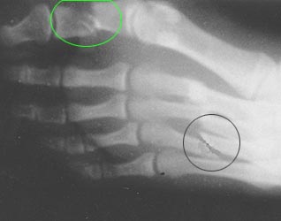
사진 240. 넷째 중족골 골절와 무지 첫째 지절골 골절(◌내)
한쪽 발에 총 27개 발 뼈가 있고 그 중 한개 또는 여러 개가 동시 골절될 수 있다.
Copyright ⓒ 2011 John Sangwon Lee, M.D.. FAAP
Fractures 골절
Figure 175. Orthopedic Patient Copyright ⓒ 2011 John Sangwon Lee, M.D., FAAP
• The numerous types of fractures and symptomatic diagnosis and treatment of fractures cannot be explained in this book.
• Depending on the age, the severity of the fracture, the type and location of the fractured bone, or various other conditions, the cause, symptom, and diagnosis of a fracture may differ.
• Fractures can be treated with observational treatment, sling treatment, crutch treatment, splint treatment, cast treatment, surgical treatment, pin treatment, and several combination treatments. Normal skeleton, fractures, and dislocations as seen in pictures and X-rays
Picture 176. Lateral X-ray of the cervical spine. The cervical spine consists of 7 vertebrae (Cervical Vertebra). One cervical vertebra or several cervical vertebrae can be fractured in a safety accident. Copyright ⓒ 2011 John Sangwon Lee, M.D., FAAP
Picture 177. Anterior X-ray of the cervical spine The cervical vertebrae in cervical vertebrae consisted of 7 vertebrae. Copyright ⓒ 2011 John Sangwon Lee, M.D., FAAP
Picture 178. Lateral X-ray of the lumbar spine The lumbar vertebrae consisted of a total of 5 vertebrae (lumbar vertebrae). Copyright ⓒ 2011 John Sangwon Lee, M.D., FAAP
Picture 179. Anterior X-ray of the thoracic and lumbar spine. The thoracic vertebrae consisted of a total of 12 vertebrae (thoracic vertebrae). Copyright ⓒ 2011 John Sangwon Lee, M.D., FAAP
Picture 185. Lateral X-ray of the lumbar spine The lumbar vertebrae consisted of a total of 5 vertebrae (lumbar vertebrae). Copyright ⓒ 2011 John Sangwon Lee, M.D., FAAP
Picture 186. Lateral X-ray of the lumbar spine Copyright ⓒ 2011 John Sangwon Lee, M.D., FAAP
Photo 187. ◯My right clavicle fracture and left normal clavicle Copyright ⓒ 2011 John Sangwon Lee, M.D., FAAP
Picture 188. There are left and right clavicles in the prominent parts left and right. Copyright ⓒ 2011 John Sangwon Lee, M.D., FAAP
Photo 189. Fracture of the right clavicle The clavicle area marked with ◯ is fractured. Copyright ⓒ 2011 John Sangwon Lee, M.D., FAAP
Picture 190.◯My left clavicle fracture The shoulder joint is within ◯. Copyright ⓒ 2011 John Sangwon Lee, M.D., FAAP
Picture 193. Right shoulder joint X-ray The right shoulder joint is within ◯. a – humerus, b – clavicle, c – scapula Copyright ⓒ 2011 John Sangwon Lee, M.D., FAAP
Photo 194. ◯My left clavicle fracture Copyright ⓒ 2011 John Sangwon Lee, M.D., FAAP
Picture 195. ◯The shoulder joint and clavicle fracture The clavicle was fractured in the area indicated by ↑. a – clavicle, b – humerus, c – scapula Copyright ⓒ 2011 John Sangwon Lee, M.D., FAAP
Photo 196. ◯My shoulder joint (scapular joint) The humerus head was fractured in the area indicated by ↑. a – humerus, b – clavicle, c – scapula Copyright ⓒ 2011 John Sangwon Lee, M.D., FAAP
Picture 197. Anterior X-ray of the elbow joint (elbow joint) A-humerus, b-radius, c-ulna, medial fossa of the humerus fractured in ◌. Copyright ⓒ 2011 John Sangwon Lee, M.D., FAAP
Picture 198. Side X-ray of the normal elbow joint (elbow joint) (in ◌) a – humerus, b – radius, b – ulna Copyright ⓒ 2011 John Sangwon Lee, M.D., FAAP
Photo 199. ◯Internal humerus fracture a – humerus, b – radius, c – ulna Copyright ⓒ 2011 John Sangwon Lee, M.D., FAAP
Picture 200. ◯ My severe humerus comminuted fracture a – humerus, b – radius, c – ulna Copyright ⓒ 2011 John Sangwon Lee, M.D., FAAP
Photo 201. ◌Internal radial fracture and ulnar fracture Copyright ⓒ 2011 John Sangwon Lee, M.D., FAAP
Picture 202. Large ◌ inner radius fracture and small ◌ inner ulnar fracture Copyright ⓒ 2011 John Sangwon Lee, M.D., FAAP
Photo 203. Radial fracture and ulnar fracture (upper and lower) Copyright ⓒ 2011 John Sangwon Lee, M.D., FAAP
Photo 204. ◌Internal radial fracture and ulnar fracture Copyright ⓒ 2011 John Sangwon Lee, M.D.. FAAP
Photo 205. ◌Internal Radial Fracture Ulnar Fracture Copyright ⓒ 2011 John Sangwon Lee, M.D.. FAAP
Picture 206. ◌My radius and ulna were fractured. Copyright ⓒ 2011 John Sangwon Lee, M.D.. FAAP
Picture 207. ◌Internal radial fracture and ulnar fracture Copyright ⓒ 2011 John Sangwon Lee, M.D. FAAP
Photo 208. 0Composite fracture of inner radius and ulna Copyright ⓒ 2011 John Sangwon Lee, M.D. FAAP
Picture 209. □My radius fracture. Copyright ⓒ 2011 John Sangwon Lee, M.D.. FAAP
Picture 210. □My radius fracture. Copyright ⓒ 2011 John Sangwon Lee, M.D. FAAP
Photo 211. ◯My hand bone fracture There are 14 finger bones (finger bones) in one hand and 8 carpal bones. There are 1-5 metacarpals corresponding to each finger. One or several bones of the hand may be fractured. Fractures can occur in the epiphysis, diaphysis, and growth plate of the hand bones. Copyright ⓒ 2011 John Sangwon Lee, M.D. FAAP
Picture 212. ◯Fracture of the first finger bone of my second finger. Copyright ⓒ 2011 John Sangwon Lee, M.D. FAAP
Picture 213. ◯Fracture of the metacarpal bone of my thumb Copyright ⓒ 2011 John Sangwon Lee, M.D.. FAAP Photo 214. ◌Fracture of the first finger bone of my thumb Copyright ⓒ 2011 John Sangwon Lee, M.D. FAAP
Photo 215. ◌The joint between the metacarpal bone of my little finger and the first finger bone of the little finger was dislocated and a fracture occurred. Copyright ⓒ 2011 John Sangwon Lee, M.D. FAAP
Photo 216. ◌I fractured the joint between the metacarpal bone of my little finger and the finger bone of my little finger, and also had a dislocation. Copyright ⓒ 2011 John Sangwon Lee, M.D. FAAP
Picture 217. Femur fracture (in ◌) a – ischial bone, b – femoral head Copyright ⓒ 2011 John Sangwon Lee, M.D. FAAP
Photo 218. Tibia fracture (inner ◌) Copyright ⓒ 2011 John Sangwon Lee, M.D. FAAP
Picture 219. Knee joint (knee joint) Copyright ⓒ 2011 John Sangwon Lee, M.D. FAAP
Photo 220. ○My normal knee joint (knee joint) a – lower femur, b – patella, c – the tibia, d – fibula Copyright ⓒ 2011 John Sangwon Lee, M.D. FAAP
Picture 221. X-ray before and after knee joint a – lower femur, b – tibia, c – fibula ◌My knee joint (knee joint). Copyright ⓒ 2011 John Sangwon Lee, M.D. FAAP
Picture 222. Lateral X-ray of the knee joint a – lower femur, b – patella, c – tibia, d – fibula ◌Internal tibia fracture. Copyright ⓒ 2011 John Sangwon Lee, M.D. FAAP
Photo 223. Normal legs and feet of infants Copyright ⓒ 2011 John Sangwon Lee, M.D. FAAP
Picture 224. Anterior and posterior X-ray of the knee joint a – tibia, b – fibula, c◯ – ankle joint, d◯ – knee joint Copyright ⓒ 2011 John Sangwon Lee, M.D. FAAP
Photo 226.◌Internal tibia fracture a◯ – knee joint, b – tibia, c – fibula, d◯ – tibia fracture, e◯ – ankle joint Copyright ⓒ 2011 John Sangwon Lee, M.D. FAAP
Picture 227. Fracture of large ◌ internal tibia, small ◌ fracture of internal fibula Copyright ⓒ 2011 John Sangwon Lee, M.D. FAAP
Picture 228. X-ray picture of normal ankle (inner ◯) and anterior foot Copyright ⓒ 2011 John Sangwon Lee, M.D. FAAP
Picture 229. Normal ankle and Achilles tendon, ankle lateral X-ray a-Achilles tendon, b-fibula, c-tibia, d-talus, e-calcaneus Copyright ⓒ 2011 John Sangwon Lee, M.D. FAAP
Photo 230. Severe ankle joint fracture and dislocation a – fibula fracture, b◯ – ankle joint, c – talus, d – tibia, e – tibial head dislocation, f – calcaneus Copyright ⓒ 2011 John Sangwon Lee, M.D. FAAP
Picture 231. Ankle joint bone and lateral X-ray image of the tarsal bone a – tibia, b – fibula There are 7 tarsal bones (talus/Talus bone, calcaneus/Calcaneus bone, 3 cuneiform/Three cuneiform bones, dice/cuboid bone, navicular/navicular bone) in one foot. For reference, there are 8 carpal bones in one hand. The tarsal bone, fibula, and tibia together form the ankle joint. The tarsal bones connect upward to the tibia and the lower part of the fibula and downward to the metatarsal. The tarsal bone may be fractured. Copyright ⓒ 2011 John Sangwon Lee, M.D. FAAP
Picture 232. Lateral X-ray of the foot. The b-foot bone consists of the tarsal, metatarsal and phalanges. Copyright ⓒ 2011 John Sangwon Lee, M.D. FAAP
Photo 234. Ankle joint fracture and dislocation ◯My severe tibial fibula fractures and dislocations. Copyright ⓒ 2011 John Sangwon Lee, M.D. FAAP
Picture 235. Metatarsal bones (1, 2, 3, 4, 5) The bones in the middle of the foot are called metatarsal bones. There are 5 metatarsal bones in one foot, which are connected to the tarsal bone in the back and to the phalanges (toes) in the front. a-second sphenoid (second cuneiform), b-navicular, c-third sphenoid (third cuneiform), d-cubic bone Copyright ⓒ 2011 John Sangwon Lee, M.D. FAAP
Photo 236. The tarsal bone at the back of the foot (in ◌) Copyright ⓒ 2011 John Sangwon Lee, M.D. FAAP
Photo 237. ◌My metatarsal bones (1-5) and tarsal bones The bones in the middle of the foot (lower of the foot) are called metatarsal bones. There are 5 metatarsal bones in each foot. Metatarsal fractures may occur. Copyright ⓒ 2011 John Sangwon Lee, M.D. FAAP
Photo 238.◯My phalanges (toe bones) The bones of the toes in the toes are called phalanges. There are 14 phalanges. Fractures in the phalanges may occur. The big toe is called the thumb. Copyright ⓒ 2011 John Sangwon Lee, M.D. FAAP
Photo 239. ◯Fracture of the first phalanges of my thumb (fracture of the toes) There are a total of 27 foot bones in one foot, and one or several of them can be fractured simultaneously. Copyright ⓒ 2011 John Sangwon Lee, M.D. FAAP
Photo 240. Fracture of the fourth metatarsal and fracture of the first phalanges of the thumb (inner ◌) There are a total of 27 foot bones in one foot, and one or several of them can be fractured simultaneously. Copyright ⓒ 2011 John Sangwon Lee, M.D. FAAP
출처 및 참조 문헌 Sources and references
- NelsonTextbook of Pediatrics 22ND Ed
- The Harriet Lane Handbook 22ND Ed
- Growth and development of the children
- Red Book 32nd Ed 2021-2024
- Neonatal Resuscitation, American Academy Pediatrics
- www.drleepediatrics.com 제16권 소아청소년 정형외과 질환
- www.drleepediatrics.com제8권 소아청소년 호흡기 질환
- www.drleepediatrics.com제9권 소아청소년 소화기 질환
- www.drleepediatrics.com제10권. 소아청소년 신장 비뇨 생식기 질환
- www.drleepediatrics.com제11권. 소아청소년 심장 혈관계 질환
- www.drleepediatrics.com제12권. 소아청소년 신경 정신 질환, 행동 수면 문제
- www.drleepediatrics.com제13권. 소아청소년 혈액, 림프, 종양 질환
- www.drleepediatrics.com제14권. 소아청소년 내분비, 유전, 염색체, 대사, 희귀병
- www.drleepediatrics.com제15권. 소아청소년 알레르기, 자가 면역질환
- Red book 29th-31st edition 2021
- Nelson Text Book of Pediatrics 19th — 21st Edition
- The Johns Hopkins Hospital, The Harriet Lane Handbook, 22nd edition
-
Childhood Emergencies in the Office, Hospital and Community, American Academy of Pediatrics
-
Emergency Medical Service for Children, By Ross Lab. May 1989. p.10
-
Emergency care, harvey grant and robert murray
-
Emergency Care Transportation of Sick and Injured American Academy of Orthopaedic Surgeons
-
Emergency Pediatrics A Guide to Ambulatory Care, Roger M. Barkin, Peter Rosen
-
Immediate care of the acutely ill and injured, Hugh E. Stephenson, Jr
-
The Critically Ill Child, Diagnosis and Management, Edited by Clement A. Smith
-
Emergency Medical Services for Children: The Role of the Primary Care Provider, America Academy of Pediatrics
-
Quick Reference To Pediatric Emergencies , Delmer J. Pascoe, M.D., Moses Grossman, M.D. with 26 contributors
-
Manual of Emergency Care ]
-
응급환자관리 정담미디어
-
소아가정간호백과–부모도 반의사가 되어야 한다, 이상원
-
Neonatal Resuscitation American heart Association
-
Neonatology Jeffrey J.Pomerance, C. Joan Richardson
-
Pediatric Resuscitation Pediatric Clinics of North America, Stephen M. Schexnayder, M.D.
-
Pediatric Critical Care, Pediatric Clinics of North America, James P. Orlowski, M.D.
-
Preparation for Birth. Berverly Savage and Dianna Smith
- Infectious disease of children, Saul Krugman, Samuel L Katz, Ann A. Gerhon, Catherine Wilfert
-
The Harriet Lane Handbook 19th Edition
-
소아과학 대한교과서
-
제1권 소아청소년 응급의료 참조문헌과 출처
-
Other
Copyright ⓒ 2015 John Sangwon Lee, MD., FAAP
“부모도 반의사가 되어야 한다”-내용은 여러분들의 의사로부터 얻은 정보와 진료를 대신할 수 없습니다.
“The information contained in this publication should not be used as a substitute for the medical care and advice of your doctor. There may be variations in treatment that your doctor may recommend based on individual facts and circumstances. “Parental education is the best medicine.”